Review
. 2020 May 27:11:776.
doi: 10.3389/fimmu.2020.00776. eCollection 2020.

Autoimmune Pathology in Myasthenia Gravis Disease Subtypes Is Governed by Divergent Mechanisms of Immunopathology

Affiliations
Review

Autoimmune Pathology in Myasthenia Gravis Disease Subtypes Is Governed by Divergent Mechanisms of Immunopathology

Miriam L Fichtner et al. Front Immunol. .

Abstract

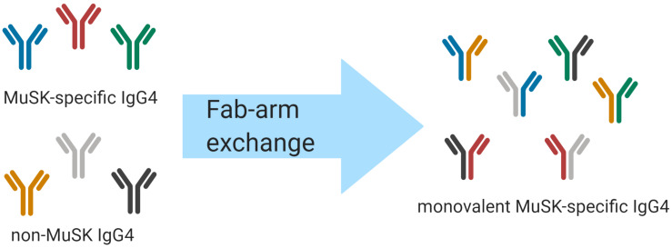
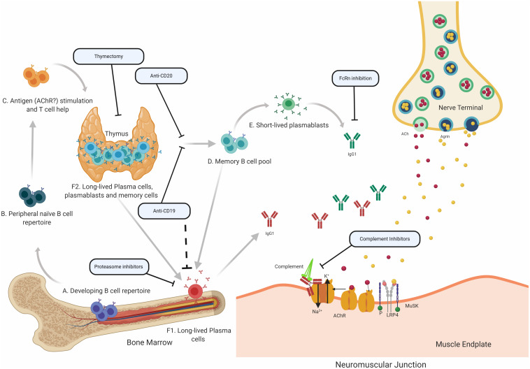
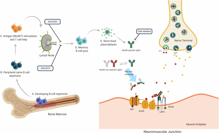
Myasthenia gravis (MG) is a prototypical autoantibody mediated disease. The autoantibodies in MG target structures within the neuromuscular junction (NMJ), thus affecting neuromuscular transmission. The major disease subtypes of autoimmune MG are defined by their antigenic target. The most common target of pathogenic autoantibodies in MG is the nicotinic acetylcholine receptor (AChR), followed by muscle-specific kinase (MuSK) and lipoprotein receptor-related protein 4 (LRP4). MG patients present with similar symptoms independent of the underlying subtype of disease, while the immunopathology is remarkably distinct. Here we highlight these distinct immune mechanisms that describe both the B cell- and autoantibody-mediated pathogenesis by comparing AChR and MuSK MG subtypes. In our discussion of the AChR subtype, we focus on the role of long-lived plasma cells in the production of pathogenic autoantibodies, the IgG1 subclass mediated pathology, and contributions of complement. The similarities underlying the immunopathology of AChR MG and neuromyelitis optica (NMO) are highlighted. In contrast, MuSK MG is caused by autoantibody production by short-lived plasmablasts. MuSK MG autoantibodies are mainly of the IgG4 subclass which can undergo Fab-arm exchange (FAE), a process unique to this subclass. In FAE IgG4, molecules can dissociate into two halves and recombine with other half IgG4 molecules resulting in bispecific antibodies. Similarities between MuSK MG and other IgG4-mediated autoimmune diseases, including pemphigus vulgaris (PV) and chronic inflammatory demyelinating polyneuropathy (CIDP), are highlighted. Finally, the immunological distinctions are emphasized through presentation of biological therapeutics that provide clinical benefit depending on the MG disease subtype.

Keywords: MuSK; AChR; B cells; B lymphocytes; autoantibodies; autoimmunity; immunopathology; myasthenia gravis.


Figures

FIGURE 1
FIGURE 1
Schematic of IgG4 Fab-arm exchange in MuSK MG. Human IgG4 antibodies can participate in a process termed Fab-arm exchange. In the MuSK autoantibody subtype of MG, MuSK-specific IgG4 autoantibodies can undergo Fab-arm exchange with other circulating IgG4 antibodies. The antibodies that were formerly divalent – with two identical antigen binding sites – become monovalent and bispecific after a heavy and light chain pair is switched with a heavy and light chain pair of another antibody. The process is thought to be critical for the development of pathogenic autoantibodies in MuSK MG.
FIGURE 2
FIGURE 2
Speculative mechanisms of AChR MG immunopathology. The proposed mechanistic path to autoantibody production in AChR MG begins with naïve B cells (Steps A and B), which likely encounter self-antigen(s) and receive T cell help in the thymus (C). They can then differentiate into autoantibody specific memory B cells (D), which can be activated into antibody-secreting short-lived plasmablasts (E) or antibody-secreting long-lived plasma cells (F), which reside in the bone marrow (F1) and may also be present in the thymus (F2) of some patients with AChR MG. It is thought that long-lived plasma cells in the bone marrow and thymus make major contributions to AChR autoantibody production. Autoantibodies migrate to the NMJ where they bind to the AChR hindering the neuromuscular transmission by directly interrupting acetylcholine signaling at the AChR. Most of the antibodies are of the IgG1 subclass which can induce the complement cascade. Several therapeutic strategies target different parts of this process in AChR MG. Thymectomy is thought to directly remove autoantibody-producing B cell and other pathogenic cell subsets. B cell depletion, mediated by anti-CD20 antibodies is thought to remove autoreactive B cells, which includes memory cells and a subset of plasmablasts. Anti-CD19 antibodies can additionally target further subsets of plasmablasts and subsets of plasma cells. Proteasome inhibitors target plasma cells and may target the disease-causing long-lived plasma cells more efficiently. FcRn inhibitors increase the elimination of circulating IgGs, which is expected to also reduce the levels of pathogenic autoantibodies. Inhibiting complement activity, with complement inhibitors, results in the disruption of the pathogenic effector functions mediated by AChR autoantibodies.
FIGURE 3
FIGURE 3
Speculative mechanisms of MuSK MG immunopathology. The proposed mechanistic path to autoantibody production in MuSK MG begins with naïve B cells (Steps A and B), which likely encounter self-antigen(s) and receive T cell help in the lymphoid tissue (C). They then differentiate into memory B cells (D) and antibody-secreting plasmablasts (E). Most autoantibodies in MuSK MG are of the IgG4 subclass. Antibodies of the IgG4 subclass can undergo the process of Fab-arm exchange with other antibodies of the IgG4 subclass. Consequently, the divalent mono-specific MuSK autoantibodies become monovalent bispecific autoantibodies. These autoantibodies migrate to the neuromuscular junction where they bind to MuSK hindering the neuromuscular transmission by blocking the LRP4 and MuSK pathway which is important for the clustering of the AChR. Several therapeutic strategies target different parts of this process in MuSK MG. B cell depletion by anti-CD20 antibodies is thought to remove B cells expressing CD20 which includes memory cells and a subset of plasmablasts. Anti CD19 antibodies can additionally target further subsets of plasmablasts. FcRn inhibitors increase the elimination of circulating IgGs, which is expected to also reduce the levels of pathogenic autoantibodies.

Similar articles

Cited by

References

    1. Gilhus NE, Skeie GO, Romi F, Lazaridis K, Zisimopoulou P, Tzartos S. Myasthenia gravis-autoantibody characteristics and their implications for therapy. Nat Rev Neurol. (2016) 12:259–68. 10.1038/nrneurol.2016.44 - DOI - PubMed
    1. Vincent A. Unravelling the pathogenesis of myasthenia gravis. Nat Rev Immunol. (2002) 2:797–804. 10.1038/nri916 - DOI - PubMed
    1. Vincent A, Beeson D, Lang B. Molecular targets for autoimmune and genetic disorders of neuromuscular transmission. Eur J Biochem. (2000) 267:6717–28. 10.1046/j.1432-1033.2000.01785.x - DOI - PubMed
    1. Hoch W, McConville J, Helms S, Newsom-Davis J, Melms A, Vincent A. Auto-antibodies to the receptor tyrosine kinase MuSK in patients with myasthenia gravis without acetylcholine receptor antibodies. Nat Med. (2001) 7:365–8. 10.1038/85520 - DOI - PubMed
    1. Zisimopoulou P, Evangelakou P, Tzartos J, Lazaridis K, Zouvelou V, Mantegazza R, et al. A comprehensive analysis of the epidemiology and clinical characteristics of anti-LRP4 in myasthenia gravis. J Autoimmun. (2014) 52:139–45. 10.1016/j.jaut.2013.12.004 - DOI - PubMed

Publication types