Spinal dural arteriovenous fistula masquerading as subdural hematoma
- PMID: 32547829
- PMCID: PMC7294149
- DOI: 10.25259/SNI_160_2020
Spinal dural arteriovenous fistula masquerading as subdural hematoma
Abstract
Background: This case highlights an angiographically occult spinal dural AVF presenting with a spinal subdural hematoma. While rare, it is important that clinicians be aware of this potential etiology of subdural hematomas before evacuation.
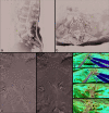
Case description: A 79-year-old female presented with acute lumbar pain, paraparesis, and a T10 sensory level loss. The MRI showed lower cord displacement due to curvilinear/triangular enhancement along the right side of the canal at the T12-L1 level. The lumbar MRA, craniospinal CTA, and multivessel spinal angiogram were unremarkable. A decompressive exploratory laminectomy revealed a subdural hematoma that contained blood products of different ages, and a large arterialized vein exiting near the right L1 nerve root sheath. The fistula was coagulated and sectioned. Postoperatively, the patient regained normal function.
Conclusion: Symptomatic subdural thoracolumbar hemorrhages from SDAVF are very rare. Here, we report a patient with an acute paraparesis and T10 sensory level attributed to an SDAVF and subdural hematoma. Despite negative diagnostic studies, even including spinal angiography, the patient underwent surgical intervention and successful occlusion of the SDAVF.
Keywords: Angiographically occult; Arteriovenous fistula; Subdural hematoma; Vascular malformation.
Copyright: © 2020 Surgical Neurology International.
Conflict of interest statement
There are no conflicts of interest.
Figures

References
-
- Cenzato M, Versari P, Righi C, Simionato F, Casali C, Giovanelli M. Spinal dural arteriovenous fistulae: Analysis of outcome in relation to pretreatment indicators. Neurosurgery. 2004;55:815–22. - PubMed
-
- Han PP, Theodore N, Porter RW, Detwiler PW, Lawton MT, Spetzler RF. Subdural hematoma from a Type I spinal arteriovenous malformation. Case report. J Neurosurg. 1999;90:255–7. - PubMed
-
- Koch C, Gottschalk S, Giese A. Dural arteriovenous fistula of the lumbar spine presenting with subarachnoid hemorrhage. Case report and review of the literature. J Neurosurg. 2004;100:385–91. - PubMed
-
- Minami M, Hanakita J, Takahashi T, Kitahama Y, Onoue S, Kino T, et al. Spinal dural arteriovenous fistula with hematomyelia caused by intraparenchymal varix of draining vein. Spine J. 2009;9:e15–9. - PubMed
-
- Spetzler RF, Detwiler PW, Riina HA, Porter RW. Modified classification of spinal cord vascular lesions. J Neurosurg. 2002;96:145–56. - PubMed
Publication types
LinkOut - more resources
Full Text Sources
Research Materials
