

Myxofibrosarcoma of the scalp with difficult preoperative diagnosis: A case report and review of the literature
- PMID: 32548167
- PMCID: PMC7281033
- DOI: 10.12998/wjcc.v8.i11.2350
Myxofibrosarcoma of the scalp with difficult preoperative diagnosis: A case report and review of the literature
Abstract
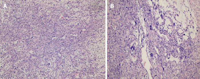
Background: A myxofibrosarcoma (MFS) is a malignant fibroblastic tumor that tends to occur in the lower and upper extremities. The reported incidence of head and neck MFSs is extremely rare. We report a 46-year-old male with "a neoplasm in the scalp" who was hospitalized and diagnosed with an MFS (highly malignant with massive necrotic lesions) based on histologic and immunohistochemistry evaluations. The magnetic resonance imaging manifestations did not demonstrate the "tail sign" mentioned in several studies, which resulted in a great challenge to establish an imaging diagnosis. The treatment plan is closely associated with the anatomic location and histologic grade, and more importantly, aggressive surgery and adjuvant radiotherapy may be helpful. Hence, we report the case and share some valuable information about the disease.
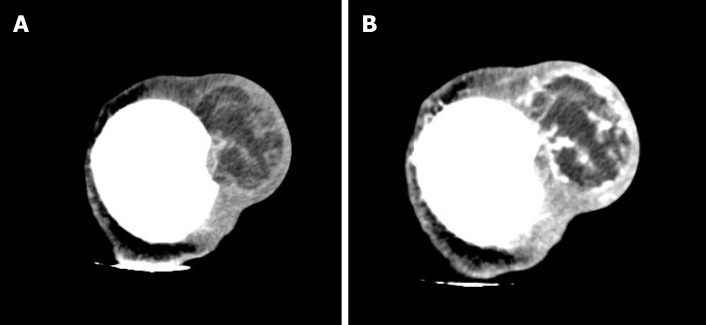
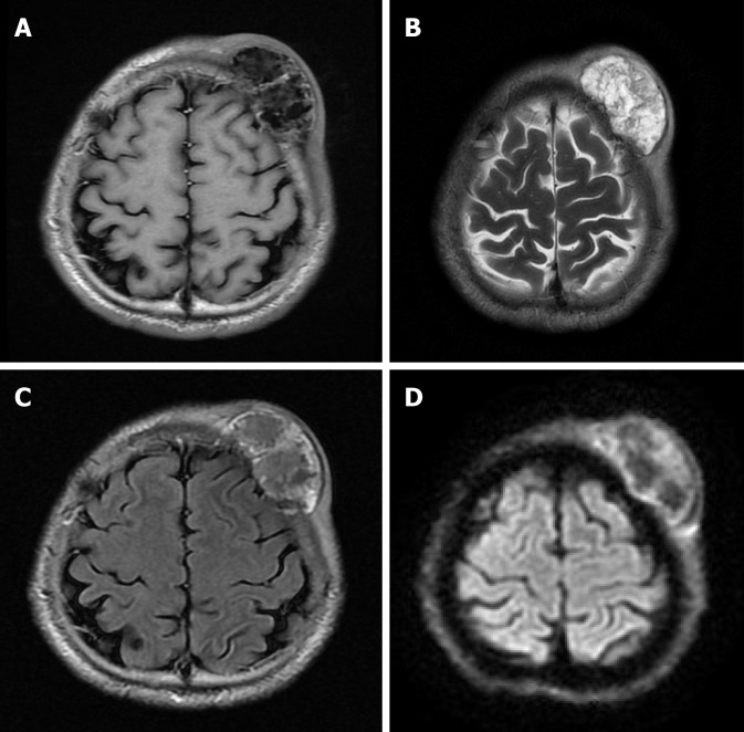
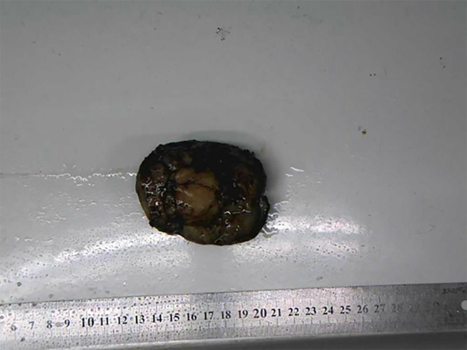
Case summary: A 46-year-old male with "a neoplasm in the scalp for 6 mo" was hospitalized. Initially, the tumor was about the size of a soybean, without algesia or ulceration. The patient ignored the growth, did not seek treatment, and thus, did not receive treatment. Recently, the tumor increased to the size of an egg; there was no bleeding or algesia. His family history was unremarkable. No abnormalities were found upon laboratory testing, including routine hematologic, biochemistry, and tumor markers. Computed tomography showed an ovoid mass (6.25 cm × 3.29 cm × 3.09 cm in size) in the left frontal scalp with low density intermingled with equidense strips in adjacent areas of the scalp. Magnetic resonance imaging revealed a lesion with an irregular surface and an approximate size of 3.55 cm × 6.34 cm in the left frontal region, with clear boundaries and visible separation. Adjacent areas of the skull were damaged and the dura mater was involved. Contrast enhancement showed an uneven enhancement pattern. Surgery was performed and postoperative adjuvant radiotherapy was administered to avoid recurrence or metastasis. The post-operative pathologic diagnosis confirmed an MFS. A repeat computed tomography scan showed no local recurrence or distant metastasis 19 mo post-operatively.
Conclusion: The case reported herein of MFS was demonstrated in an extremely rare location on the scalp and had atypical magnetic resonance imaging findings, which serves as a reminder to radiologists of the possibility of this diagnosis to assist in clinical treatment. Given the special anatomic location and the high malignant potential of this rare tumor, combined surgical and adjuvant radiotherapy should be considered to avoid local recurrence and distant metastasis. The significance of regular follow-up is strongly recommended to improve the long-term survival rate.
Keywords: Case report; Magnetic resonance imaging; Malignant fibrous histiocytoma; Myxofibrosarcoma; Scalp; Treatment.
©The Author(s) 2020. Published by Baishideng Publishing Group Inc. All rights reserved.
Conflict of interest statement
Conflict-of-interest statement: The authors have no conflicts of interest to declare.
Figures
References
-
- Pujari A, Ali MJ, Honavar SG, Mittal R, Naik M. Orbital myxofibrosarcoma: a clinicopathologic correlation of an extremely rare tumor. Ophthalmic Plast Reconstr Surg. 2014;30:e111–e113. - PubMed
-
- Nakahara S, Uemura H, Kurita T, Suzuki M, Fujii T, Tomita Y, Yoshino K. A case of myxofibrosarcoma of the maxilla with difficulty in preoperative diagnosis. Int J Clin Oncol. 2012;17:390–394. - PubMed
Publication types
LinkOut - more resources
Full Text Sources
