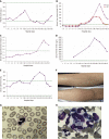
Hemophagocytic syndrome as a complication of acute pancreatitis: A case report
- PMID: 32548169
- PMCID: PMC7281054
- DOI: 10.12998/wjcc.v8.i11.2364
Hemophagocytic syndrome as a complication of acute pancreatitis: A case report
Abstract
Background: Haemophagocytic syndrome (HPS) is rarely seen in patients with acute pancreatitis (AP). HPS as a complication of AP in patients without any previous history has not been elucidated.
Case summary: A 46-year-old man was admitted for symptom of persistent abdominal pain, nausea, and vomiting for 2 d after heavy drinking. During hospital stay, he suddenly developed skin rash and a secondary fever. The laboratory findings revealed progressive pancytopenia, abnormal hepatic tests, and elevation of serum triglyceride, ferritin, and lactate dehydrogenase levels. However, apparent bacterial or viral infections were not detected. He was also possibly related to autoimmune diseases because of positive expression of various autoimmune antibodies and no remarkable past history. Finally, the bone marrow examination showed a histiocytic reactive growth and prominent hemophagocytosis, which resulted in a diagnosis of HPS. Unexpectedly, the patient responded well to the immunosuppressive therapy.
Conclusion: HPS is a very rare extrapancreatic manifestation of AP. The diagnosis relies on bone marrow examination and immunosuppressive therapy is effective. For AP with skin changes, the possibility of HPS should be considered during clinical work.
Keywords: Acute pancreatitis; Haemophagocytic syndrome; Immunosuppressive therapy.
©The Author(s) 2020. Published by Baishideng Publishing Group Inc. All rights reserved.
Conflict of interest statement
Conflict-of-interest statement: The authors declare that they have no conflict of interest.
Figures


References
-
- Reiner AP, Spivak JL. Hematophagic histiocytosis. A report of 23 new patients and a review of the literature. Medicine (Baltimore) 1988;67:369–388. - PubMed
-
- Kanaji S, Okuma K, Tokumitsu Y, Yoshizawa S, Nakamura M, Niho Y. Hemophagocytic syndrome associated with fulminant ulcerative colitis and presumed acute pancreatitis. Am J Gastroenterol. 1998;93:1956–1959. - PubMed
-
- Ishii E, Ohga S, Imashuku S, Kimura N, Ueda I, Morimoto A, Yamamoto K, Yasukawa M. Review of hemophagocytic lymphohistiocytosis (HLH) in children with focus on Japanese experiences. Crit Rev Oncol Hematol. 2005;53:209–223. - PubMed
-
- Janka GE, Lehmberg K. Hemophagocytic syndromes--an update. Blood Rev. 2014;28:135–142. - PubMed
Publication types
LinkOut - more resources
Full Text Sources
Miscellaneous

