Review
. 2020 May 11;4(6):114-146.
doi: 10.15698/cst2020.06.221.

Mitochondria in cancer

Affiliations
Review

Mitochondria in cancer

Debora Grasso et al. Cell Stress. .

Abstract

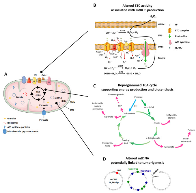
The rediscovery and reinterpretation of the Warburg effect in the year 2000 occulted for almost a decade the key functions exerted by mitochondria in cancer cells. Until recent times, the scientific community indeed focused on constitutive glycolysis as a hallmark of cancer cells, which it is not, largely ignoring the contribution of mitochondria to the malignancy of oxidative and glycolytic cancer cells, being Warburgian or merely adapted to hypoxia. In this review, we highlight that mitochondria are not only powerhouses in some cancer cells, but also dynamic regulators of life, death, proliferation, motion and stemness in other types of cancer cells. Similar to the cells that host them, mitochondria are capable to adapt to tumoral conditions, and probably to evolve to 'oncogenic mitochondria' capable of transferring malignant capacities to recipient cells. In the wider quest of metabolic modulators of cancer, treatments have already been identified targeting mitochondria in cancer cells, but the field is still in infancy.

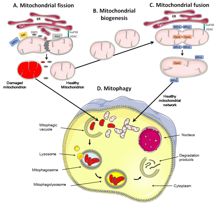
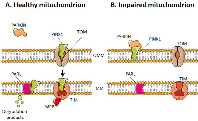
Keywords: apoptosis; mitochondrial biogenesis; mitophagy; oxidative phosphorylation (OXPHOS); reactive oxygen species (ROS); tricarboxylic acid (TCA) cycle; tumor metabolism.


Conflict of interest statement

Conflict of interest: The authors declare no conflict of interest.

Figures

Figure 1
Figure 1. FIGURE 1: Cancer is associated with alterations of mitochondrial functions.
(A) Mitochondria in normal and in cancer cells are composed of three compartments. They are separated from the cell cytosol by an outer membrane (OMM), an intermembrane space (IMS), and an inner membrane (IMM) that forms invaginations called "crests". The IMM delimitates the mitochondrial matrix, a gelatinous material containing mitochondrial DNA (mtDNA), granules, ribosomes and ATP synthase particles. The mitochondrial matrix hosts the tricarboxylic acid (TCA) cycle, while the IMM hosts the electron transport chain (ETC). (B) In highly metabolically active or hypoxic cancer cells much more than in normal cells under normal conditions, electrons escape during mitochondrial electron transport at Complexes I and III generate superoxide (O2-) from oxygen (O2) in both the IMS and the matrix. O2- is immediately dismutated to H2O2 either spontaneously or under the catalysis of superoxide dismutases SOD1 (in the IMS) or SOD2 (in the matrix). In the matrix, H2O2 can be neutralized by glutathione (GSH). It can also signal to the cytosol. (C) In cancer cells, the TCA cycle not only serve to produce reducing equivalents to fuel the ETC (green arrows), but also to generate biosynthetic intermediates that are necessary for cell proliferation (pink arrows). The most important anaplerotic reaction produces oxaloacetate directly from pyruvate, and is catalyzed by pyruvate carboxylase (PC) (blue arrow). Oxaloacetate can further be converted to phosphoenolpyruvate (PEP) by PEP carboxykinase (PC), contributing to gluconeogenesis. (D) Mitochondrial DNA (mtDNA) variations, including single nucleotide polymorphisms (SNPs), maternally inherited haplotypes and deletions have been studied for their association with cancer. Among these, only large mtDNA deletions seem to be associated with malignancies. Cyt c - cytochrome c; Gpx - glutathione peroxidase; Q - coenzyme Q10.
Figure 2
Figure 2. FIGURE 2: The arsenal of mitochondrial antioxidant defenses comprises the thioredoxin and peroxiredoxin pathways.
The image depicts redox reactions catalyzed by the thioredoxin and peroxiredoxin systems, comprising thioredoxin reductases (TrxR, of which ThxR2 is expressed in mitochondria), thioredoxins (Trx), peroxiredoxins (Prx) and NADPH. The electron source is NADPH, which mostly originates from the pentose phosphate pathway (PPP). Oxidized thioredoxins (Trx-S2) are reduced by NADPH and selenoenzymes TrxRs. Electrons are sequentially transferred from NADPH to FAD, to the N-terminal redox active disulfide in one subunit of TrxR, and, finally, to the C-terminal active site of another subunit. Reduced thioredoxins (Trx-SH2) catalyze disulfide bond reduction in many proteins, including Prxs, thus ensuring oxidative damage repair in proteins as well as H2O2 detoxification.
Figure 3
Figure 3. FIGURE 3: A high mitochondrial turnover rate is characteristic of many cancer cells.
Mitochondrial quality control involves fission and mitophagy to eliminate defective mitochondria, whereas repopulation and functionalization depends on mitochondrial biogenesis and fusion. (A) During fission, the mitochondrion is marked and anchored by the endoplasmic reticulum (ER), notably through the binding of inositol 1,4,5-trisphosphate receptor (InsP3R) at the ER surface to voltage-dependent anion channel (VDAC) at the mitochondrial surface. This leads to the recruitment of dynamin-related protein 1 (DRP1), mitochondrial receptor protein 1 (Fis1), mitochondrial fission factor (Mff) and mitochondrial dynamic proteins (MIDs), allowing oligomerization and constriction to yield two daughter mitochondria. (B) During mitochondrial biogenesis, a mitochondrion self-replicates. (C) During fusion, mitofusins Mfn1 and Mfn2 are located on the outer mitochondrial membrane, allowing the exchange of calcium for signaling and creating antiparallel connections between two fusing mitochondria. Optic atrophy 1 (Opa1) together with Mnf1 participate in the fusion of the inner mitochondrial membrane. Fusion allows the formation of mitochondrial networks. (D) The mitophagic process consists in the engulfment of damaged mitochondria in a vacuole, called ‘mitophagic vacuole'. The subsequent fusion of the mitophagosome with lysosomes, forming a mitophagolysosome, allows the degradation of mitochondria in macromolecules that are delivered to the cytosol. Mitophagy can be non-selective or selective, using canonical and non-canonical pathways. It prevents the accumulation of damaged mitochondria that could harm or even kill the cell if apoptosis and/or the production of reactive oxygen species would derail.
Figure 4
Figure 4. FIGURE 4: Dysfunctional mitochondria are targeted to mitophagy.
(A) Healthy mitochondria have a polarized outer mitochondrial membrane (OMM), which allows PTEN-induced kinase 1 (PINK1) to cross the membrane and be degraded by presenilin-associated rhomboid-like protein (PARL) at the inner mitochondrial membrane (IMM). (B) Impaired mitochondria, instead, have a depolarized OMM, which hinders the entry of PINK1 and, therefore, its degradation. PINK1 can thus bind to parkin to initiate mitophagy. MPP - matrix-processing peptidase; TIM - translocase of inner mitochondrial membrane; TOM - translocase of outer mitochondrial membrane.

References

    1. Warburg O, Wind F, Negelein E. The metabolism of tumors in the body. J Gen Physiol. 1927;8(6):519–530. doi: 10.1085/jgp.8.6.519. - DOI - PMC - PubMed
    1. Reichert AS, Neupert W. Mitochondriomics or what makes us breathe. Trends Genet. 2004;20(11):555–562. doi: 10.1016/j.tig.2004.08.012. - DOI - PubMed
    1. Wallace DC. Mitochondrial diseases in man and mouse. Science. 1999;283(5407):1482–1488. doi: 10.1126/science.283.5407.1482. - DOI - PubMed
    1. Nomoto S, Yamashita K, Koshikawa K, Nakao A, Sidransky D. Mitochondrial D-loop mutations as clonal markers in multicentric hepatocellular carcinoma and plasma. Clin Cancer Res. 2002;8(2):481–487. - PubMed
    1. Eng C, Kiuru M, Fernandez MJ, Aaltonen LA. A role for mitochondrial enzymes in inherited neoplasia and beyond. Nat Rev Cancer. 2003;3(3):193–202. doi: 10.1038/nrc1013. - DOI - PubMed

LinkOut - more resources