

Coagulation factor XIII activity predicts left ventricular remodelling after acute myocardial infarction
- PMID: 32548915
- PMCID: PMC7524135
- DOI: 10.1002/ehf2.12774
Coagulation factor XIII activity predicts left ventricular remodelling after acute myocardial infarction
Abstract
Aims: Acute myocardial infarction (MI) is the major cause of chronic heart failure. The activity of blood coagulation factor XIII (FXIIIa) plays an important role in rodents as a healing factor after MI, whereas its role in healing and remodelling processes in humans remains unclear. We prospectively evaluated the relevance of FXIIIa after acute MI as a potential early prognostic marker for adequate healing.
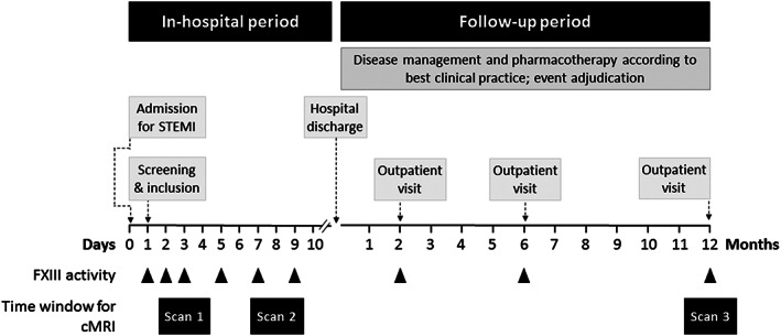
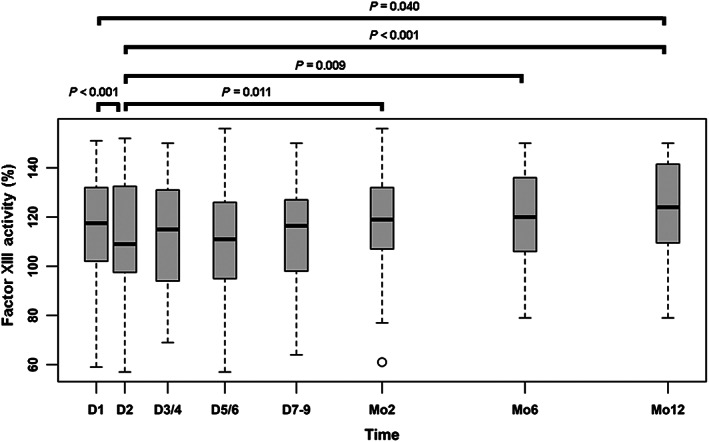
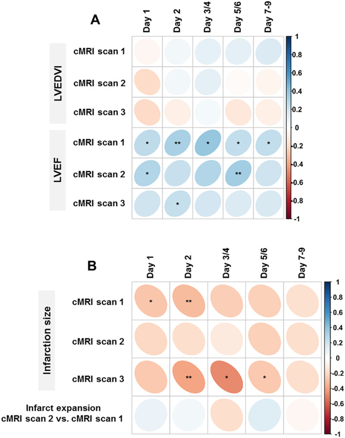
Methods and results: This monocentric prospective cohort study investigated cardiac remodelling in patients with ST-elevation MI and followed them up for 1 year. Serum FXIIIa was serially assessed during the first 9 days after MI and after 2, 6, and 12 months. Cardiac magnetic resonance imaging was performed within 4 days after MI (Scan 1), after 7 to 9 days (Scan 2), and after 12 months (Scan 3). The FXIII valine-to-leucine (V34L) single-nucleotide polymorphism rs5985 was genotyped. One hundred forty-six patients were investigated (mean age 58 ± 11 years, 13% women). Median FXIIIa was 118% (quartiles, 102-132%) and dropped to a trough on the second day after MI: 109% (98-109%; P < 0.001). FXIIIa recovered slowly over time, reaching the baseline level after 2 to 6 months and surpassed baseline levels only after 12 months: 124% (110-142%). The development of FXIIIa after MI was independent of the genotype. FXIIIa on Day 2 was strongly and inversely associated with the relative size of MI in Scan 1 (Spearman's ρ = -0.31; P = 0.01) and Scan 3 (ρ = -0.39; P < 0.01) and positively associated with left ventricular ejection fraction: ρ = 0.32 (P < 0.01) and ρ = 0.24 (P = 0.04), respectively.
Conclusions: FXIII activity after MI is highly dynamic, exhibiting a significant decline in the early healing period, with reconstitution 6 months later. Depressed FXIIIa early after MI predicted a greater size of MI and lower left ventricular ejection fraction after 1 year. The clinical relevance of these findings awaits to be tested in a randomized trial.
Keywords: Blood coagulation factor XIII; Cardiac magnetic resonance imaging; Healing and remodelling processes; ST-elevation myocardial infarction.
© 2020 The Authors. ESC Heart Failure published by John Wiley & Sons Ltd on behalf of the European Society of Cardiology.
Conflict of interest statement
None declared.
Figures
Similar articles
-
Factor XIIIA-V34L and factor XIIIB-H95R gene variants: effects on survival in myocardial infarction patients.Mol Med. 2007 Jan-Feb;13(1-2):112-20. doi: 10.2119/2006-00049.Gemmati. Mol Med. 2007. PMID: 17515963 Free PMC article.
-
Transglutaminase activity in acute infarcts predicts healing outcome and left ventricular remodelling: implications for FXIII therapy and antithrombin use in myocardial infarction.Eur Heart J. 2008 Feb;29(4):445-54. doi: 10.1093/eurheartj/ehm558. Eur Heart J. 2008. PMID: 18276618
-
F13A1 Gene Variant (V34L) and Residual Circulating FXIIIA Levels Predict Short- and Long-Term Mortality in Acute Myocardial Infarction after Coronary Angioplasty.Int J Mol Sci. 2018 Sep 14;19(9):2766. doi: 10.3390/ijms19092766. Int J Mol Sci. 2018. PMID: 30223472 Free PMC article.
-
Clinical aspects of left ventricular diastolic function assessed by Doppler echocardiography following acute myocardial infarction.Dan Med Bull. 2001 Nov;48(4):199-210. Dan Med Bull. 2001. PMID: 11767125 Review.
-
The effects of different initiation time of exercise training on left ventricular remodeling and cardiopulmonary rehabilitation in patients with left ventricular dysfunction after myocardial infarction.Disabil Rehabil. 2016;38(3):268-76. doi: 10.3109/09638288.2015.1036174. Epub 2015 Apr 17. Disabil Rehabil. 2016. PMID: 25885667 Review.
Cited by
-
Sustained Increase in Serum Glial Fibrillary Acidic Protein after First ST-Elevation Myocardial Infarction.Int J Mol Sci. 2022 Sep 7;23(18):10304. doi: 10.3390/ijms231810304. Int J Mol Sci. 2022. PMID: 36142218 Free PMC article.
-
Machine Learning Analysis of the Cerebrovascular Thrombi Proteome in Human Ischemic Stroke: An Exploratory Study.Front Neurol. 2020 Nov 5;11:575376. doi: 10.3389/fneur.2020.575376. eCollection 2020. Front Neurol. 2020. PMID: 33240201 Free PMC article.
-
Differential Role of Factor XIII in Acute Myocardial Infarction and Ischemic Stroke.Biomedicines. 2024 Feb 22;12(3):497. doi: 10.3390/biomedicines12030497. Biomedicines. 2024. PMID: 38540111 Free PMC article. Review.
-
Interaction between Acute Hepatic Injury and Early Coagulation Dysfunction on Mortality in Patients with Acute Myocardial Infarction.J Clin Med. 2023 Feb 15;12(4):1534. doi: 10.3390/jcm12041534. J Clin Med. 2023. PMID: 36836066 Free PMC article.
-
Use of serial changes in biomarkers vs. baseline levels to predict left ventricular remodelling after STEMI.ESC Heart Fail. 2023 Feb;10(1):432-441. doi: 10.1002/ehf2.14204. Epub 2022 Oct 21. ESC Heart Fail. 2023. PMID: 36271665 Free PMC article.
References
-
- Gaudron P, Eilles C, Kugler I, Ertl G. Progressive left ventricular dysfunction and remodeling after myocardial infarction. Potential mechanisms and early predictors. Circulation 1993; 87: 755–763. - PubMed
-
- Ertl G, Frantz S. Healing after myocardial infarction. Cardiovasc Res 2005; 66: 22–32. - PubMed
-
- Ertl G, Frantz S. Wound model of myocardial infarction. Am J Physiol Heart Circ Physiol 2005; 288: H981–H983. - PubMed
-
- Jackson BM, Gorman JH, Moainie SL, Guy TS, Narula N, Narula J, John‐Sutton MG, Edmunds LH Jr, Gorman RC. Extension of borderzone myocardium in postinfarction dilated cardiomyopathy. J Am Coll Cardiol 2002; 40: 1160–1167 discussion 1168‐71. - PubMed
Publication types
MeSH terms
Substances
Grants and funding
LinkOut - more resources
Full Text Sources
Medical
