. 2020 Aug;333(7):511-525.
doi: 10.1002/jez.2393. Epub 2020 Jun 17.

Is aquaporin-3 involved in water-permeability changes in the killifish during hypoxia and normoxic recovery, in freshwater or seawater?

Affiliations

Is aquaporin-3 involved in water-permeability changes in the killifish during hypoxia and normoxic recovery, in freshwater or seawater?

Ilan M Ruhr et al. J Exp Zool A Ecol Integr Physiol. 2020 Aug.

Abstract

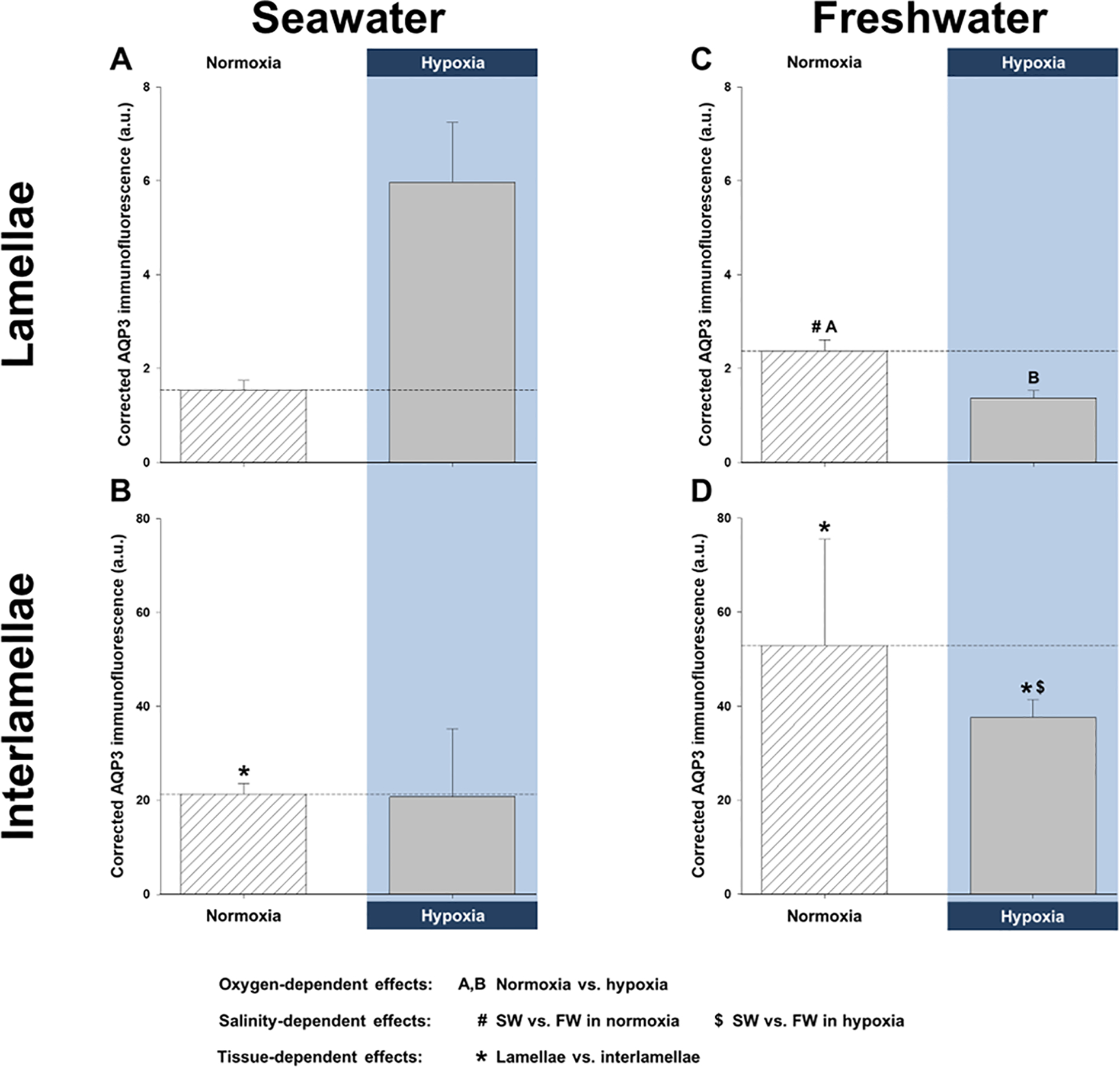
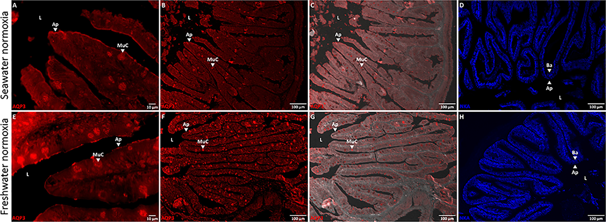
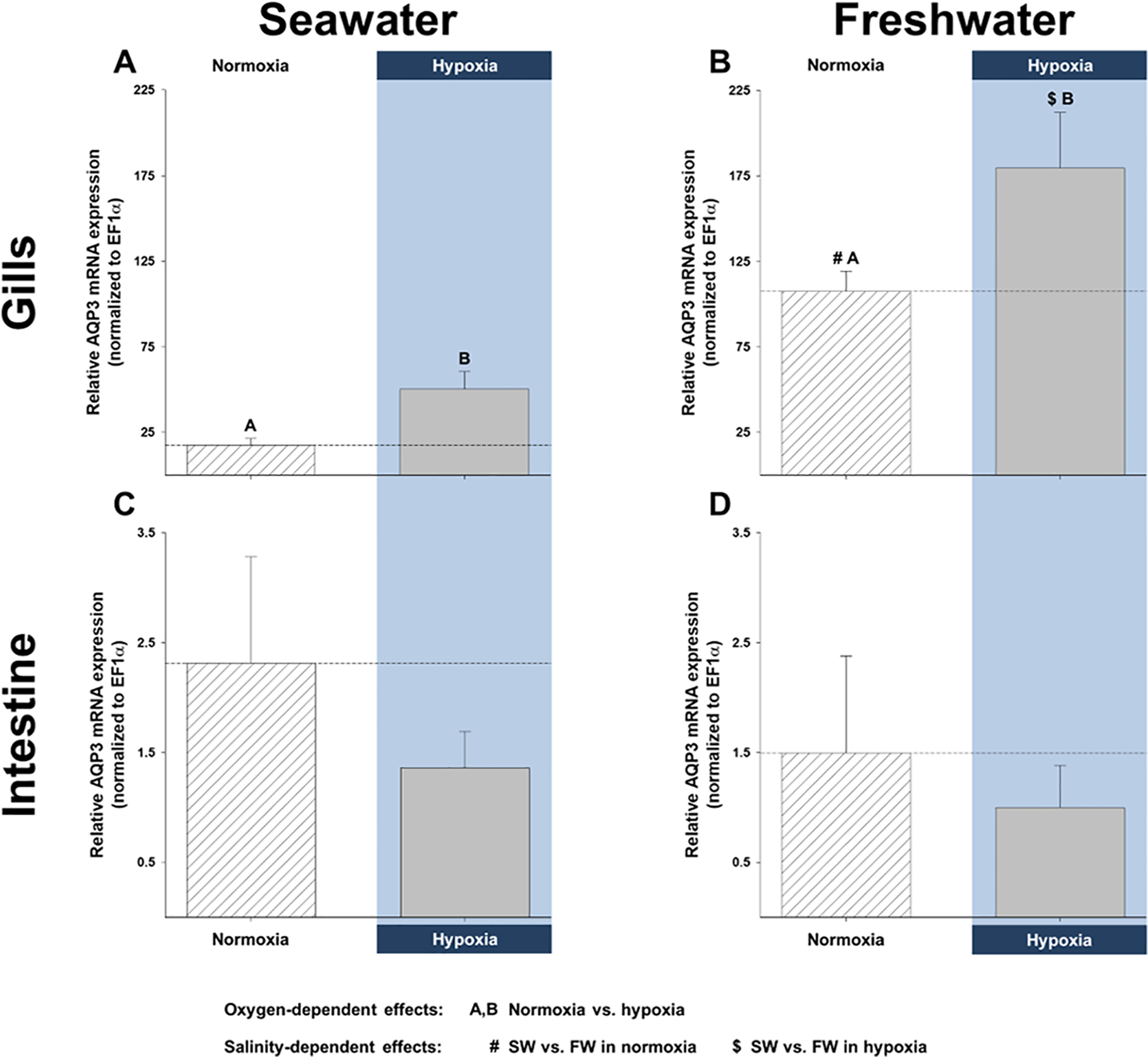
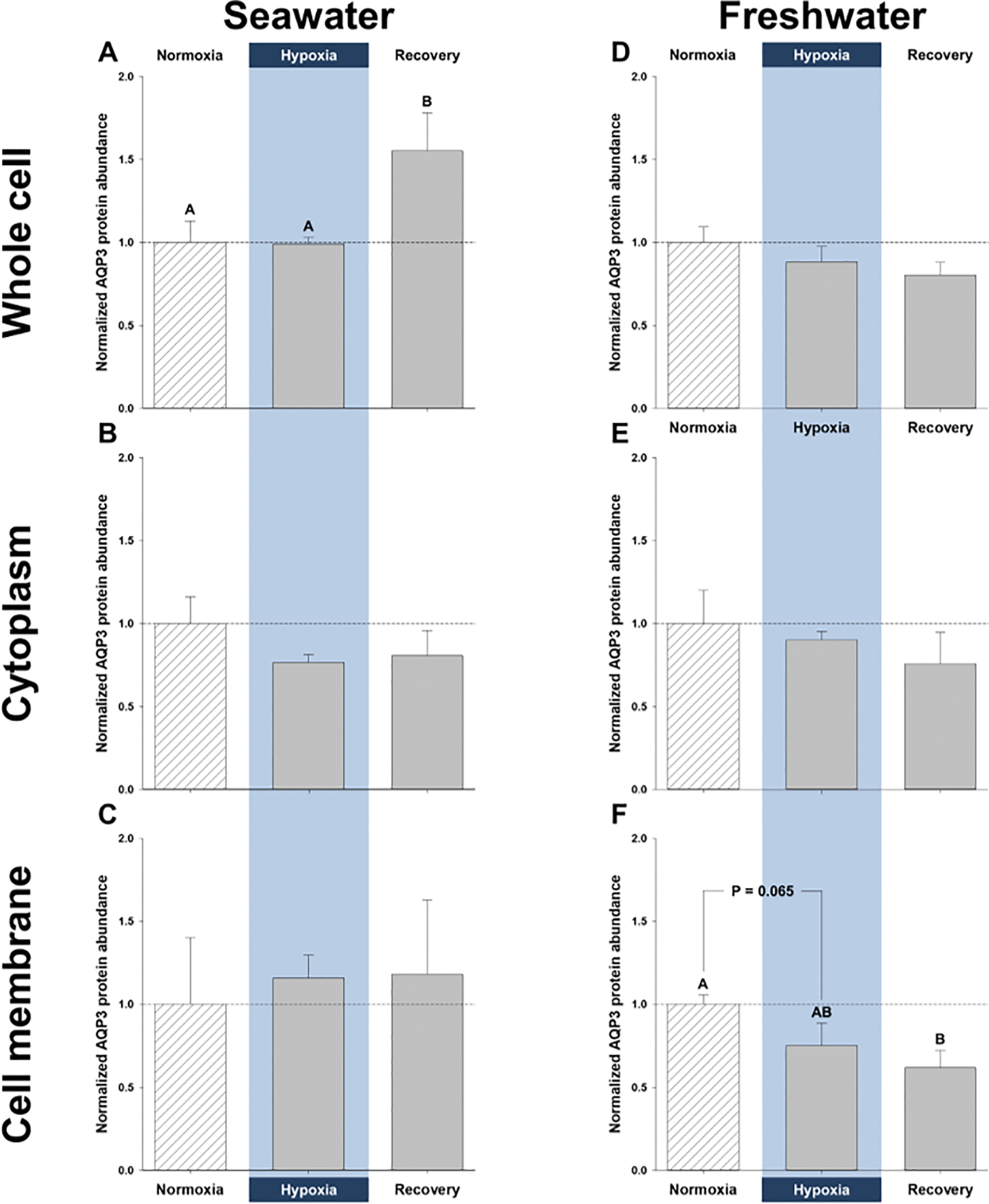
Aquaporins are the predominant water-transporting proteins in vertebrates, but only a handful of studies have investigated aquaporin function in fish, particularly in mediating water permeability during salinity challenges. Even less is known about aquaporin function in hypoxia (low oxygen), which can profoundly affect gill function. Fish deprived of oxygen typically enlarge gill surface area and shrink the water-to-blood diffusion distance, to facilitate oxygen uptake into the bloodstream. However, these alterations to gill morphology can result in unfavorable water and ion fluxes. Thus, there exists an osmorespiratory compromise, whereby fish must try to balance high branchial gas exchange with low ion and water permeability. Furthermore, the gills of seawater and freshwater teleosts have substantially different functions with respect to osmotic and ion fluxes; consequently, hypoxia can have very different effects according to the salinity of the environment. The purpose of this study was to determine what role aquaporins play in water permeability in the hypoxia-tolerant euryhaline common killifish (Fundulus heteroclitus), in two important osmoregulatory organs-the gills and intestine. Using immunofluorescence, we localized aquaporin-3 (AQP3) protein to the basolateral and apical membranes of ionocytes and enterocytes, respectively. Although hypoxia increased branchial AQP3 messenger-RNA expression in seawater and freshwater, protein abundance did not correlate. Indeed, hypoxia did not alter AQP3 protein abundance in seawater and reduced it in the cell membranes of freshwater gills. Together, these observations suggest killifish AQP3 contributes to reduced diffusive water flux during hypoxia and normoxic recovery in freshwater and facilitates intestinal permeability in seawater and freshwater.

Keywords: Fundulus heteroclitus; gills; immunofluorescence; intestine; mRNA; osmorespiratory compromise; oxygen deprivation; permeability; protein; western blot.


Figures

Figure 1.
Figure 1.. Effects of salinity and dissolved oxygen on branchial protein abundance of killifish aquaporin-3 (AQP3).
Killifish were acclimated to either seawater or freshwater, and subjected to normoxia (the control), 3 h hypoxia (10% O2 saturation), or normoxic recovery, after which the gills were excised. Three fractions were isolated from the gills (whole cell, cytoplasm, and cell membrane) and western blots were run to determine AQP3 protein abundance. (A) Whole-cell, (B) cytoplasmic, and (C) cell-membrane gill fractions of seawater-acclimated fish. (D) Whole-cell, (E) cytoplasmic, and (F) cell-membrane fractions of freshwater-acclimated fish. Data were normalized to normoxic protein levels and are shown as means ± SEM (N = 4–5). Significant differences in AQP3 protein abundance between normoxia, hypoxia, and recovery are indicated for oxygen-dependent effects, by dissimilar upper-case letters (A, B), when P ≤ 0.05. Note that seawater and freshwater samples were run on different gels, so cannot be compared statistically.
Figure 2.
Figure 2.. Effects of salinity and dissolved oxygen on branchial and intestinal mRNA expression of killifish aquaporin-3 (AQP3).
Killifish were acclimated to either seawater or freshwater, and subjected to normoxia (the control) or 3 h hypoxia (10% O2 saturation), after which the gills and intestine were excised. Relative AQP3 mRNA expression was determined by real-time, quantitative PCR and normalized by calibrating with EF1α. mRNA expression in (A) seawater and (B) freshwater of the gills and in (C) seawater and (D) freshwater of the intestine. Data are shown as means ± SEM (N = 6 fish per treatment). Significant differences in AQP3 mRNA expression are indicated for oxygen-dependent effects, by dissimilar upper-case letters (A, B), and for salinity-dependent effects, by pound signs (#) and dollar symbols ($), when P ≤ 0.05, as shown in the figure legend (tissue-dependent effects were not analyzed). Although not indicated, freshwater resulted in significantly higher total branchial AQP3 mRNA expression.
Figure 3.
Figure 3.. Effects of salinity and dissolved oxygen on killifish aquaporin-3 (AQP3) protein immunofluorescence in the lamellae and interlamellae of the gills.
Killifish were acclimated to either seawater or freshwater, and subjected to normoxia (the control) or 3 h hypoxia (10% O2 saturation), after which the gills were excised. Relative corrected AQP3 immunofluorescence [in arbitrary units (a.u.)] measured in the (A) lamellae and (B) interlamellae of seawater-acclimated fish and in the (C) lamellae and (D) interlamellae of freshwater-acclimated fish. Note the difference in scale between Panels A and C (lamellae) versus B and D (interlamellae), demonstrating that the majority of AQP3 immunofluorescence is in the interlamellar regions of the gill. Data are shown as means ± SEM (N = 6 different fish per treatment). Significant differences in AQP3 immunofluorescence are indicated for oxygen-dependent effects, by dissimilar upper-case letters (A, B), for salinity-dependent effects, by pound signs (#) and dollar symbols ($), and for tissue-dependent effects, by asterisks (*), when P ≤ 0.05, as shown in the figure legend. Although not indicated, freshwater resulted in significantly higher total branchial AQP3 immunofluorescence than in seawater, and total AQP3 immunofluorescence was significantly lower in hypoxic vs. normoxic freshwater.
Figure 4.
Figure 4.. Representative immunofluorescent images of aquaporin-3 (AQP3), the cystic fibrosis transmembrane conductance regulator (CFTR), and the Na+/K+-ATPase (NKA) in killifish gills.
Killifish were acclimated to either seawater or freshwater, and subjected to normoxia (the control) or 3 h hypoxia (10% O2 saturation), after which the gills were excised. Immunohistochemistry was used to identify the membrane localization of AQP3 (in red), CFTR (in green), and NKA (in blue), with immunofluorescent antibodies, in seawater normoxia (A, B, and C) or hypoxia (D, E, and F) and in freshwater normoxia (G, H, and I) or hypoxia (J, K, and L). All immunofluorescent images of AQP3/CFTR are in 60x and those of NKA are in 40x. AQP3 immunofluorescence generally shared the same distribution as NKA; AQP3 was localized to the basolateral membranes (Ba; example shown on Panel C) of the cells of both the gill lamellae (L), and interlamellar regions (IL) of the filaments (F). AQP3 might also occur on the apical membranes at the outer borders of the lamellae. CFTR immunofluorescence was poor in the freshwater gills, but was clearly discernible in the apical membrane of the interlamellae of seawater gills. NKA immunofluorescence was expressed on the basolateral membranes of the cells of the lamella and interlamellae under all treatments, and was particularly prominent in the interlamellar regions of the seawater gill under normoxia. Ionocytes (inset on Panel C) were apparent in the interlamellar regions of seawater gills under normoxia and hypoxia and were distinguishable by an apical crypt (ApC; Panels C and F) expressing CFTR and the basolateral membrane expressing NKA.
Figure 5.
Figure 5.. Immunofluorescent images of aquaporin-3 (AQP3) and the Na+/K+-ATPase (NKA) in the killifish intestine.
Killifish were acclimated to either seawater or freshwater and immunohistochemistry was used to identify the membrane localization of AQP3 (in red) and NKA (in blue), with immunofluorescent antibodies. (A and E) 60x, (B and F) 10x, and (C and G) 10x brightfield-overlaid images of AQP3 immunofluorescence in the killifish intestine. (D and H) 10x images of NKA immunofluorescence in the killifish intestine. AQP3 immunofluorescence is localized to the apical membrane (Ap) and mucus cells (MuC) of the enterocytes – facing the intestinal lumen (L) – and is distributed differently than NKA immunofluorescence, which is exclusively basolateral (Ba).

Similar articles

Cited by

References

    1. Baronio D, Martinez D, Fiori CZ, Bambini-Junior V, Forgiarini LF, Pase da Rosa D, Kim LJ, and Cerski MR (2013). Altered aquaporins in the brains of mice submitted to intermittent hypoxia model of sleep apnea. Respir Physiol Neurobiol, 185(2), 217–221. doi:10.1016/j.resp.2012.10.012 - DOI - PubMed
    1. Bœuf G and Payan P (2001). How should salinity influence fish growth? Comp Biochem Physiol C Toxicol Pharmacol, 130(4), 411–423. doi:10.1016/s1532-0456(01)00268-x - DOI - PubMed
    1. Breves JP, Inokuchi M, Yamaguchi Y, Seale AP, Hunt BL, Watanabe S, Lerner DT, Kaneko T, and Grau EG (2016). Hormonal regulation of aquaporin 3: opposing actions of prolactin and cortisol in tilapia gill. J Endocrinol, 230(3), 325–337. doi:10.1530/JOE-16-0162 - DOI - PubMed
    1. Breves JP, Starling JA, Popovski CM, Doud JM, and Tipsmark CK (2020). Salinity-dependent expression of ncc2 in opercular epithelium and gill of mummichog (Fundulus heteroclitus). J Comp Physiol B, 190(2), 219–230. doi:10.1007/s00360-020-01260-x - DOI - PubMed
    1. Brunelli E, Mauceri A, Salvatore F, Giannetto A, Maisano M, and Tripepi S (2010). Localization of aquaporin 1 and 3 in the gills of the rainbow wrasse, Coris julis. Acta Histochem, 112(3), 251–258. doi:10.1016/j.acthis.2008.11.030 - DOI - PubMed

Publication types