. 2020 Jul;9(14):e2000181.
doi: 10.1002/adhm.202000181. Epub 2020 Jun 17.

Targeted Heating of Mitochondria Greatly Augments Nanoparticle-Mediated Cancer Chemotherapy

Affiliations

Targeted Heating of Mitochondria Greatly Augments Nanoparticle-Mediated Cancer Chemotherapy

Jiangsheng Xu et al. Adv Healthc Mater. 2020 Jul.

Abstract

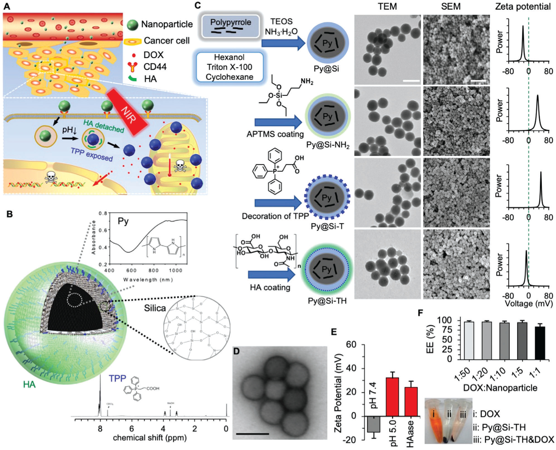
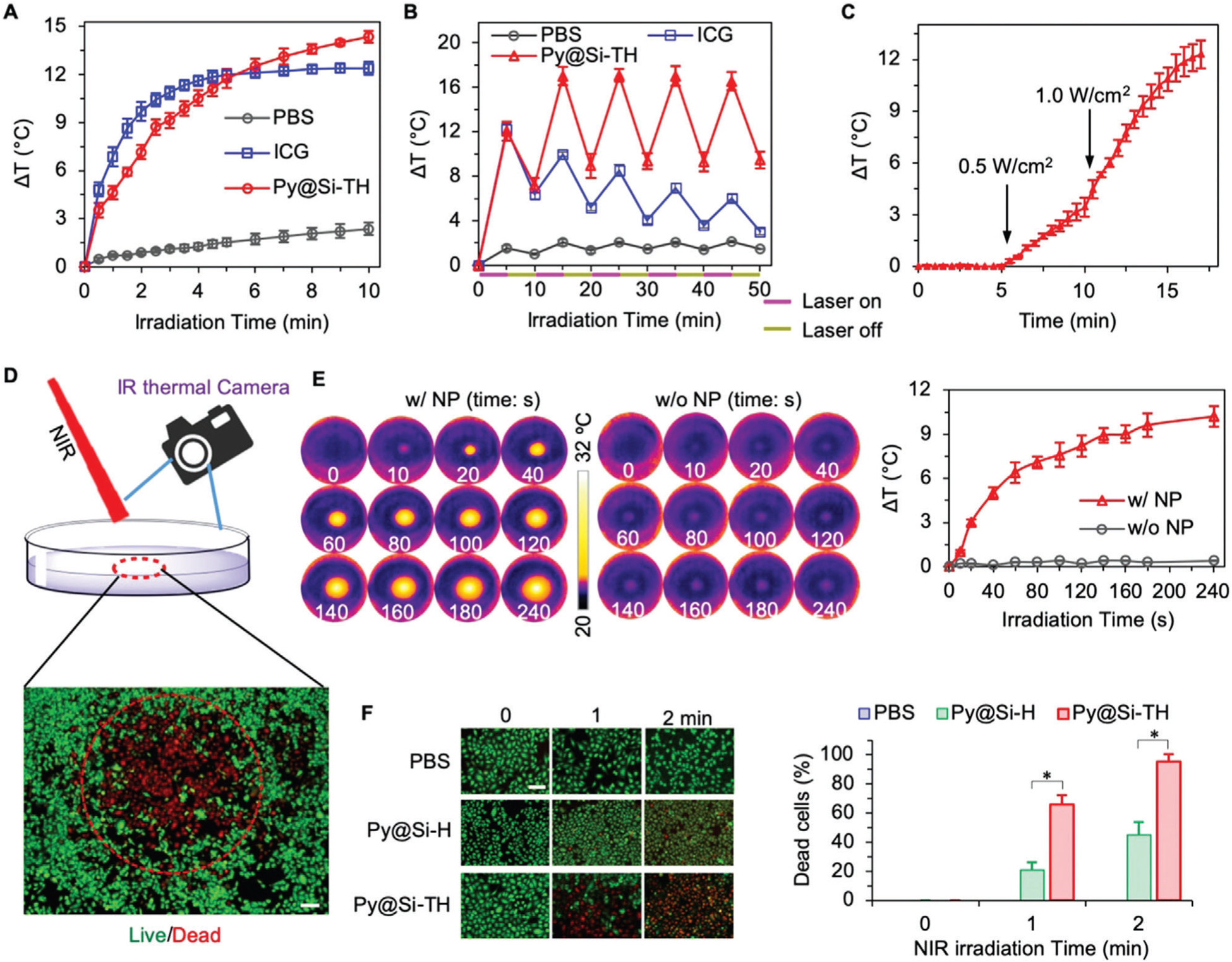
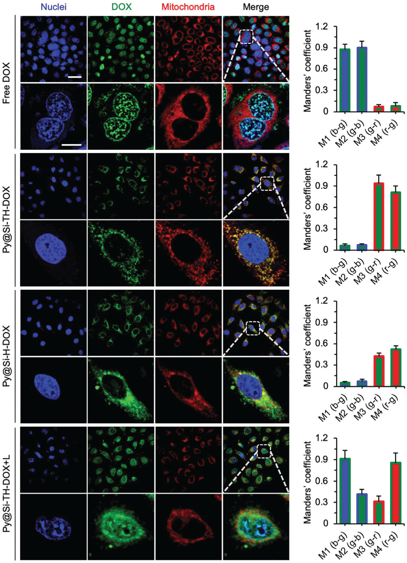
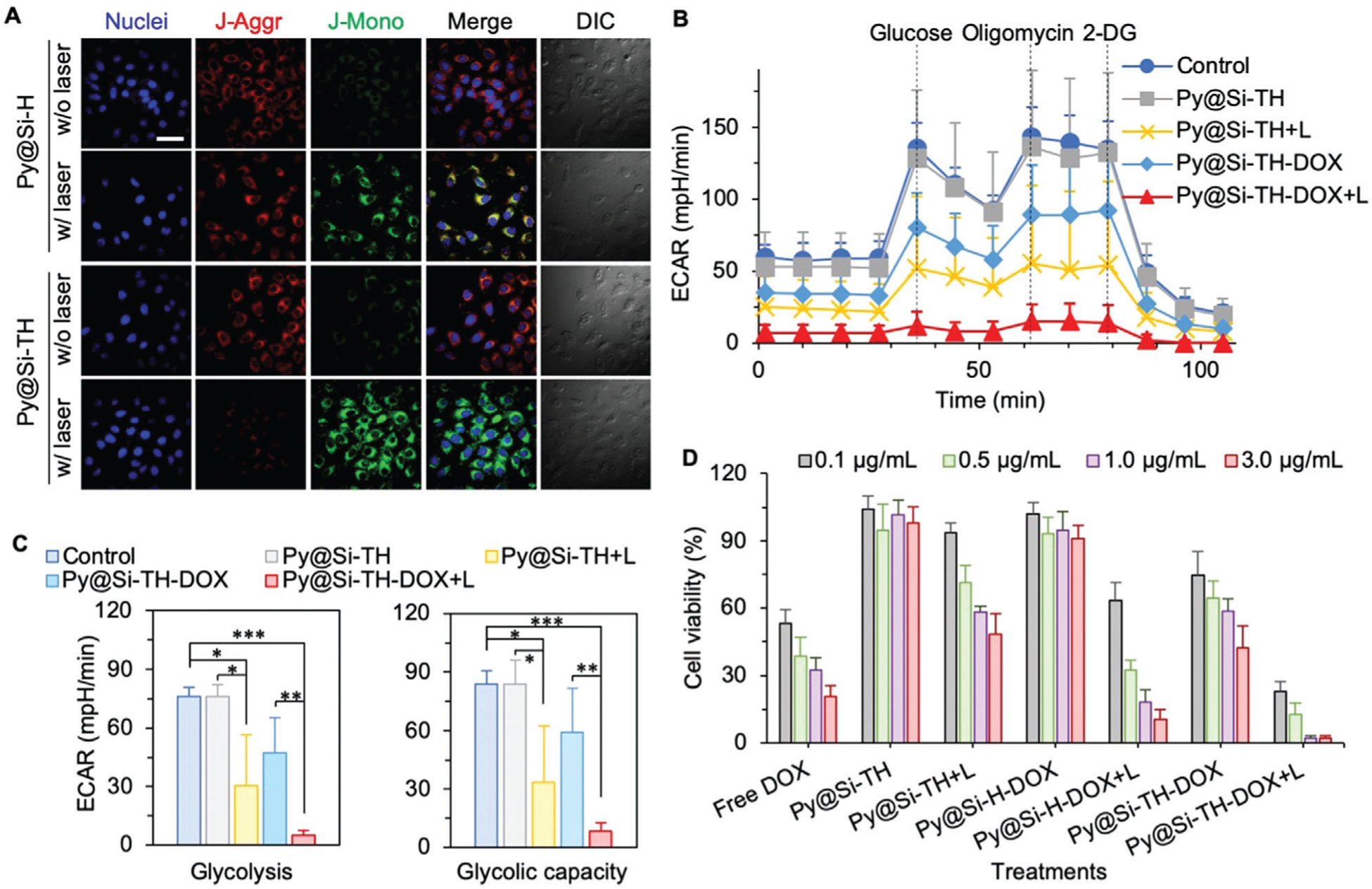
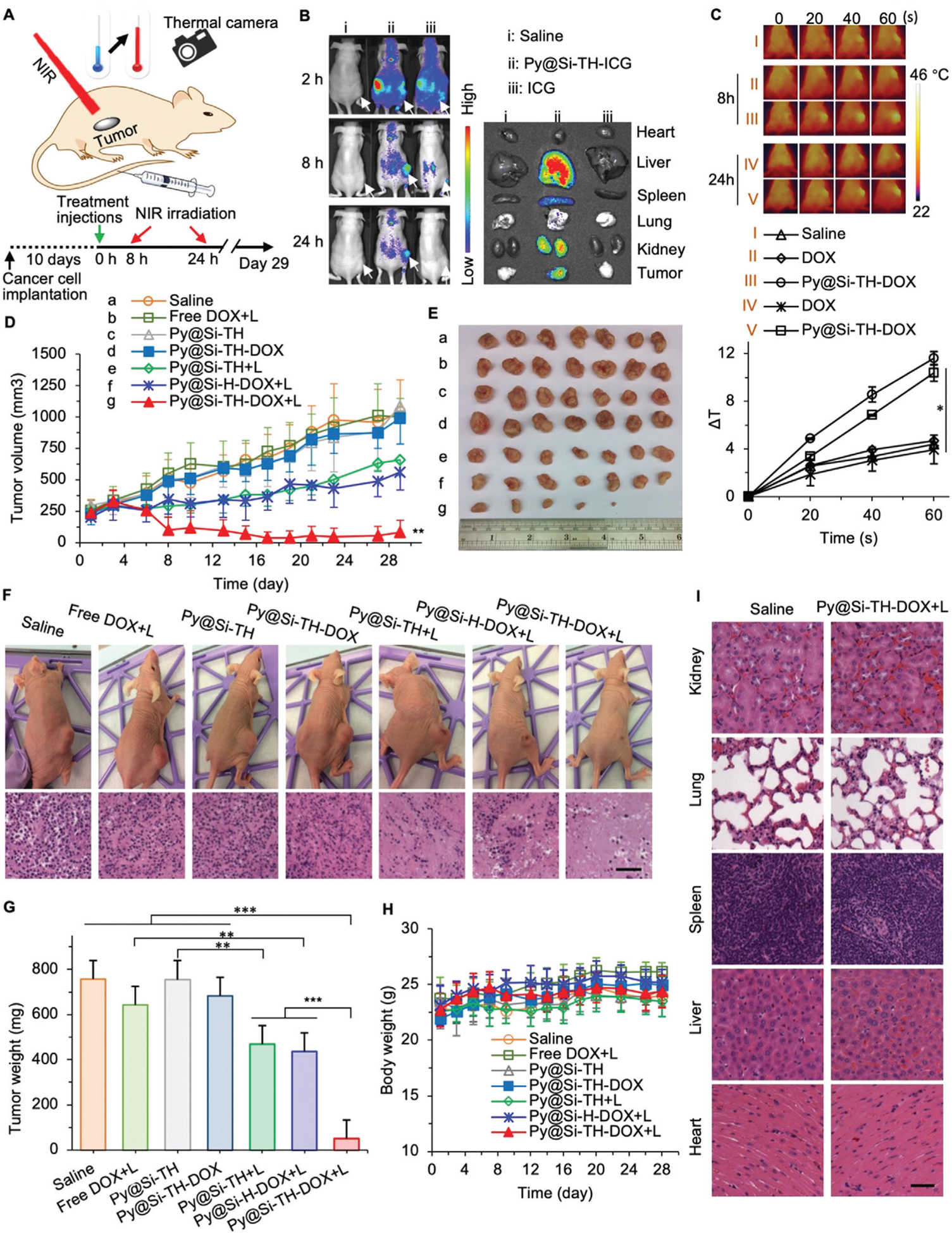
Cancer is the second leading cause of mortality globally. Various nanoparticles have been developed to improve the efficacy and safety of chemotherapy, photothermal therapy, and their combination for treating cancer. However, most of the existing nanoparticles are low in both subcellular precision and drug loading content (<≈5%), and the effect of targeted heating of subcellular organelles on the enhancement of chemotherapy has not been well explored. Here, a hybrid Py@Si-TH nanoparticle is reported to first target cancer cells overexpressed with the variant CD44 via its natural ligand HA on the outermost surface of the nanoparticle before cellular uptake, and then target mitochondria after they are taken up inside cells. In addition, the nanoparticle is ultraefficient for encapsulating doxorubicin hydrochloride (DOX) to form Py@Si-TH-DOX nanoparticle. The encapsulation efficiency is ≈100% at the commonly used low feeding ratio of 1:20 (DOX:empty nanoparticle), and >80% at an ultrahigh feeding ratio of 1:1. In combination with near infrared (NIR, 808 nm) laser irradiation, the tumor weight in the Py@Si-TH-DOX treatment group is 8.5 times less than that in the Py@Si-H-DOX (i.e., DOX-laden nanoparticles without mitochondrial targeting) group, suggesting targeted heating of mitochondria is a valuable strategy for enhancing chemotherapy to combat cancer.

Keywords: conductive polymers; drug delivery; mitochondria targeting; photothermal therapy; ultrahigh anti-cancer efficiency.


Conflict of interest statement

Conflict of Interest

The authors declare no conflict of interest.

Figures

Figure 1.
Figure 1.
Synthesis and characterization of nanoparticles. A) A schematic illustration of the nanoparticle in targeting tumor via the enhanced permeability and retention (EPR) effect of tumor vasculature, cancer cells via CD44, and mitochondria via triphenylphosphonium (TPP) for chemo-photothermal therapy of cancer. B) A schematic illustration of the structure of the nanoparticle together with materials in the nanoparticles. Py: polypyrrole. HA: hyaluronic acid. C) The procedure for preparing Py-embedded silica (Py@Si) nanoparticles, modifying the Py@Si nanoparticles with (3-aminopropyl) trimethoxysilane (APTMS) to form Py@Si-NH2 nanoparticles, coating the Py@Si-NH2 nanoparticles with TPP to produce mitochondria-targeting Py@Si-T nanoparticles, and finally, coating the Py@Si-T nanoparticles with HA to produce Py@Si-TH nanoparticles. Transmission electron microscopy (TEM) and scanning electron microscopy (SEM) images were also given to show the morphology of the nanoparticles. The difference in surface zeta potential of the Py@Si, Py@Si-NH2, Py@Si-T, and Py@Si-TH nanoparticles demonstrating the successful sequential modification of the positively charged APTMS and TPP and negatively charged HA on the nanoparticle surface. Scale bar: 100 and 500 nm for the TEM and SEM images, respectively. D) TEM images of Py@Si-TH nanoparticles with negative staining showing the HA coating on the outermost surface of the nanoparticles. Scale bar: 100 nm). E) Zeta potential data showing HA detachment from Py@Si-TH nanoparticles after treatment at low pH or with hyaluronidase (HAase). All the zeta potential data were measured by redispersing the nanoparticles in deionized water at neutral pH after the treatments. F) Encapsulation efficiency (EE) of doxorubicin hydrochloride (DOX) by simply mixing it with Py@Si-TH nanoparticles at different feeding ratios (DOX:nanoparticle in weight) for 1 h. Centrifuge tubes depicting the color of DOX, Py@Si-TH nanoparticles, and mixture of DOX and Py@Si-TH nanoparticles (1:1) after centrifuging (13 000 × g), indicating the high EE of DOX with the nanoparticles at the high feeding ratio.
Figure 2.
Figure 2.
Photothermal effect of nanoparticles. A) Increase in temperature of phosphate buffered saline (PBS) and PBS suspended/dissolved with Py@Si-TH nanoparticles (30 μg mL−1) or free ICG (5 μg mL−1) upon NIR laser irradiation at 1.0 W cm−2. B) Photothermal effect of Py@Si-TH nanoparticles versus ICG in PBS with five cycles of on/off NIR laser irradiation, showing excellent photothermal stability of the nanoparticles compared to ICG. C) Laser power density dependent photothermal effect of Py@Si-TH nanoparticles, showing different temperature increasing rates from 0.5 to 1.0 W cm−2. D) A schematic diagram showing MDA-MB-231 cells in a dish treated with Py@Si-TH nanoparticles upon NIR laser irradiation. The red circle shows the area of irradiation. The local temperature change was monitored by an FLIR E6 infrared (IR) thermal camera. Cells were stained with calcein AM and propidium iodide (PI) to fluorescently visualize the live/dead information after laser irradiation. Scale bar: 200 μm. E) IR thermal images of MDA-MB-231 cells with (w/ NP) or without (w/o NP) Py@Si-TH nanoparticles upon laser irradiation, and the corresponding curves of temperature change. F) Fluorescence microscopy images showing live/dead (green/red) cells and the percentage of dead cells (the number of PI positive cells to the total number of cells) after treating MDA-MB-231 cells with PBS, Py@Si-H nanoparticles, and Py@Si-TH nanoparticles in combination with laser irradiation for 0, 1, or 2 min. Statistical significance was assessed by Student’s t-test (unpaired and two-tailed); n = 3; and scale bar: 50 μm. *p < 0.05.
Figure 3.
Figure 3.
Cell uptake of nanoparticles and their subcellular distribution. Confocal images showing cell uptake and subcellular distribution of Py@Si-TH-DOX nanoparticles compared with free DOX and Py@Si-H-DOX nanoparticles (no mitochondria targeting capability) in MDA-MB-231 cells. Cells were incubated with MitoTracker Deep Red (red) and DAPI (blue) to stain their mitochondria and nuclei, respectively. The merged and zoom-in images show effective overlap of mitochondria (red) and the Py@Si-TH-DOX nanoparticles (green), demonstrating the mitochondria-targeting capability of the nanoparticles. Such overlap is minimal for the Py@Si-H-DOX nanoparticles. Furthermore, the overlap between cell nuclei (blue) and DOX (green) is evident after NIR laser irradiation of the cells treated with Py@Si-TH-DOX nanoparticles (i.e., Py@Si-TH-DOX+L), because of the high binding affinity between DOX and the nuclear materials. This indicates the NIR laser-triggered release of DOX from the nanoparticles inside cells. These qualitative observations are confirmed by quantitative analyses of the colocalization of DOX with nuclei and mitochondria using Manders’ coefficients: M1 (b–g) denotes the fraction of nuclei/blue overlapping with DOX/green, M2 (g–b) denotes the fraction of DOX/green overlapping with nuclei/blue, M3 (g–r) denotes the fraction of DOX/green overlapping with mitochondria/red, and M4 (r–g) denotes the fraction of mitochondria/red overlapping with DOX/green. Scale bars: 20 μm and 5 μm for low and high magnification images, respectively.
Figure 4.
Figure 4.
Mitochondrial damage analyses and in vitro anticancer efficacy. A) Mitochondrial membrane potential of MDA-MB-231 cells loaded with Py@Si-TH (with mitochondria targeting) versus Py@Si-H (without mitochondria targeting) nanoparticles with or without laser irradiation, indicating enhanced mitochondrial damage with Py@Si-TH nanoparticles under NIR laser irradiation. Scale bar: 20 μm. B) Real-time extracellular acidification rate (ECAR) analysis of MDA-MB-231 cells either without any treatment (control) or after treated with Py@Si-TH, Py@Si-TH+L (L: NIR laser irradiation), Py@Si-TH-DOX, and Py@Si-TH-DOX+L. 2-DG: 2-deoxy-d-glucose. C) Comparison of glycolysis and glycolytic capacity (the summation of glycolysis and glycolytic reserve) of the cells with different treatments. Statistical significance was assessed by Student’s t-test (unpaired and two tailed); n = 3; and *p < 0.05, **p < 0.01, ***p < 0.001. D) Targeted heating of mitochondria augments chemotherapy in vitro. The MDA-MB-231 cells were greatly sensitized to nanoparticle-mediated chemotherapy after targeted heating (+L) of mitochondria.
Figure 5.
Figure 5.
In vivo tumor targeting and antitumor efficacy and safety. A) A schematic diagram of the in vivo experiment showing tumor-bearing mice were injected with various drug formulations including the Py@Si-TH-DOX nanoparticles intravenously via the tail vein followed by NIR laser irradiation (+L for some mice), together with a sketch describing the exact timing according to which the injections/treatments of the mice were performed from the implantation of cancer cells. Temperature change in the tumor area was monitored with an IR thermal camera. B) In vivo whole animal imaging of ICG fluorescence at 2, 8, and 24 h after intravenous injection of saline, Py@Si-TH-ICG nanoparticles (i.e., Py@Si-TH nanoparticles encapsulated with ICG), or free ICG, showing effective tumor targeting of the Py@Si-TH nanoparticles. The arrows indicate the locations of tumors in mice. Also shown is the ex vivo imaging of tumors and critical organs collected from the mice sacrificed at 24 h, confirming the tumor targeting capability of the Py@Si-TH nanoparticle observed with whole animal imaging. C) IR thermal images (top) of mice injected with free DOX or Py@Si-TH-DOX nanoparticles upon laser irradiation for various time periods (0–60 s) at 8 h and 24 h after the injections. Saline was used as control. The corresponding heating curves (bottom) show the significantly increased temperature in tumor for the group of Py@Si-TH-DOX upon NIR laser irradiation. Error bars represent s.d. (n = 3). D) Tumor growth and E) a photo of the tumors collected on day 29 after sacrificing the mice. F) Photos of representative mice on day 29 for the seven different treatments, together with representative images of the histology (Hematoxylin&eosin or H&E stain) of the tumors from mice for the seven treatments. G) Weight of the tumors collected after sacrificing the mice on day 29. H) Body weight of the mice with the various treatments showing no significant change for the mice with the seven treatments. I) Histological (H&E) images of five major organs in mice treated with saline or Py@Si-TH-DOX+L collected on day 29. All NIR laser irradiation was administered at 1.0 W cm−2 for 1 min. The NIR irradiation were conducted at 8 h and 24 h after intravenous administration of the various treatments. Statistical significance was assessed by Student t-test when comparing two groups (G), and one-way ANOVA with a Fisher’s least significant difference (LSD) post hoc test (C,D) or a Dunnett’s post hoc test (G) when comparing more than two groups; error bars represent s.d. (n = 7); scale bars: 50 μm; and *p < 0.05, **p < 0.01.

Similar articles

Cited by

References

    1. Mayer EL, Burstein HJ, J. Clin. Oncol 2016, 34, 3369; - PubMed
    2. Denkert C, Liedtke C, Tutt A, von Minckwitz G, Lancet 2017, 389, 2430. - PubMed
    1. Miller KD, Siegel RL, Lin CC, Mariotto AB, Kramer JL, Rowland JH, Stein KD, Alteri R, Jemal A, Cancer J Clin. 2016, 66, 271; - PubMed
    2. Wang H, Agarwal P, Zhao S, Yu J, Lu X, He X, Nat. Commun 2015, 6, 10081; - PMC - PubMed
    3. Oualla K, El-Zawahry HM, Arun B, Reuben JM, Woodward WA, Gamal El-Din H, Lim B, Mellas N, Ueno NT, Fouad TM, Ther. Adv. Med. Oncol 2017, 9, 493; - PMC - PubMed
    4. Schettini F, Giuliano M, De Placido S, Arpino G, Cancer Treat. Rev 2016, 50, 129; - PubMed
    5. Locatelli MA, Curigliano G, Eniu A, Breast Care 2017, 12, 152; - PMC - PubMed
    6. Yao H, He G, Yan S, Chen C, Song L, Rosol TJ, Deng X, Oncotarget 2017, 8, 1913; - PMC - PubMed
    7. Zeichner SB, Terawaki H, Gogineni K, Breast Cancer 2016, 10, 25. - PMC - PubMed
    1. Dean M, Fojo T, Bates S, Nat. Rev. Cancer 2005, 5, 275. - PubMed
    1. Blanco E, Shen H, Ferrari M, Nat. Biotechnol 2015, 33, 941; - PMC - PubMed
    2. Sherlock SP, Tabakman SM, Xie L, Dai H, ACS Nano 2011, 5, 1505; - PMC - PubMed
    3. Langer R, Science 249, 1990, 1527; - PubMed
    4. Farokhzad OC, Langer R, ACS Nano 2009, 3, 16; - PubMed
    5. Hare JI, Lammers T, Ashford MB, Puri S, Storm G, Barry ST, Adv. Drug Delivery Rev 2017, 108, 25; - PubMed
    6. Wilhelm S, Tavares AJ, Dai Q, Ohta S, Audet J, Dvorak HF, Chan WCW, Nat. Rev. Mater 2016, 1, 16014.
    1. Kamaly N, Yameen B, Wu J, Farokhzad OC, Chem. Rev 2016, 116, 2602; - PMC - PubMed
    2. Masoud H, Alexeev A, ACS Nano 2012, 6, 212; - PubMed
    3. Slowing II, Vivero-Escoto JL, Wu C-W, Lin VS-Y, Adv. Drug Delivery Rev 2008, 60, 1278. - PubMed

Publication types