. 2020 Jun 15;10(6):907.
doi: 10.3390/biom10060907.

Structural Basis of Specific Glucoimidazole and Mannoimidazole Binding by Os3BGlu7

Affiliations

Structural Basis of Specific Glucoimidazole and Mannoimidazole Binding by Os3BGlu7

Bodee Nutho et al. Biomolecules. .

Abstract

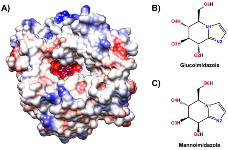
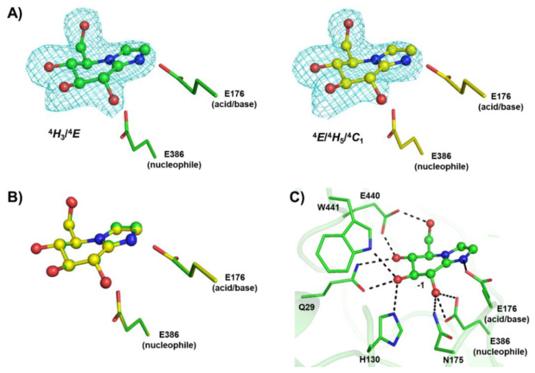
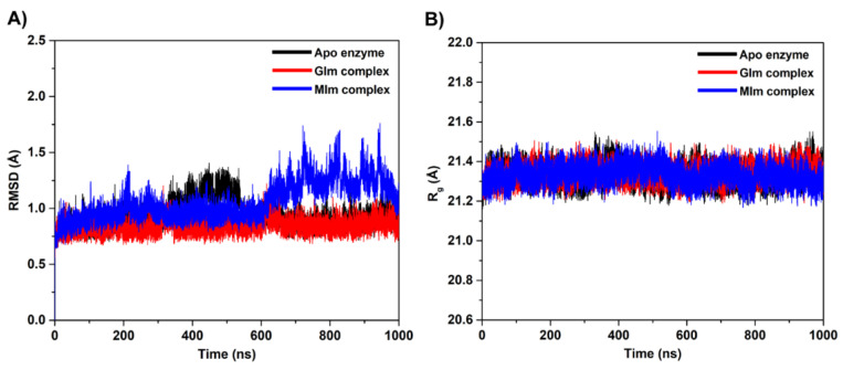
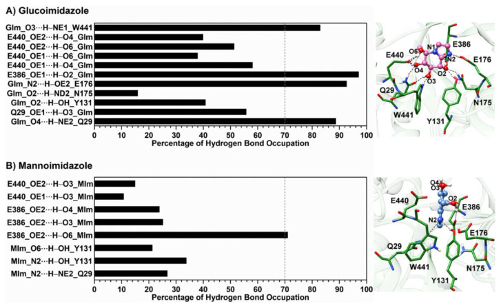
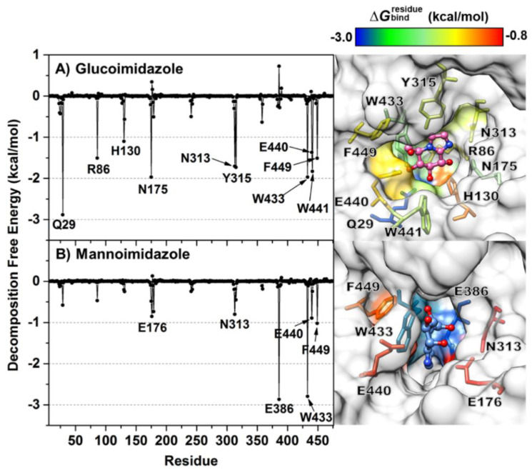
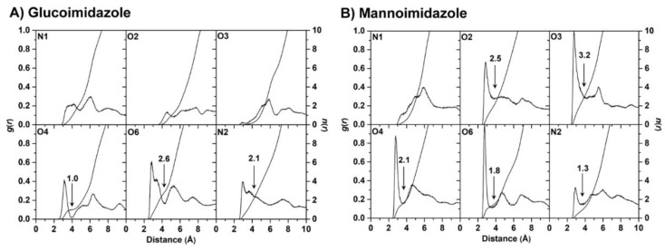
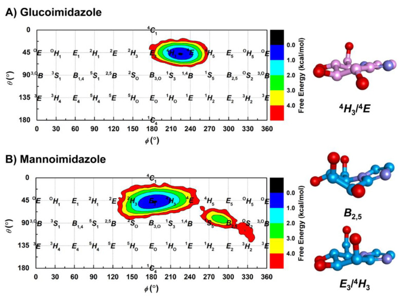
β-Glucosidases and β-mannosidases hydrolyze substrates that differ only in the epimer of the nonreducing terminal sugar moiety, but most such enzymes show a strong preference for one activity or the other. Rice Os3BGlu7 and Os7BGlu26 β-glycosidases show a less strong preference, but Os3BGlu7 and Os7BGlu26 prefer glucosides and mannosides, respectively. Previous studies of crystal structures with glucoimidazole (GIm) and mannoimidazole (MIm) complexes and metadynamic simulations suggested that Os7BGlu26 hydrolyzes mannosides via the B2,5 transition state (TS) conformation preferred for mannosides and glucosides via their preferred 4H3/4E TS conformation. However, MIm is weakly bound by both enzymes. In the present study, we found that MIm was not bound in the active site of crystallized Os3BGlu7, but GIm was tightly bound in the -1 subsite in a 4H3/4E conformation via hydrogen bonds with the surrounding residues. One-microsecond molecular dynamics simulations showed that GIm was stably bound in the Os3BGlu7 active site and the glycone-binding site with little distortion. In contrast, MIm initialized in the B2,5 conformation rapidly relaxed to a E3/4H3 conformation and moved out into a position in the entrance of the active site, where it bound more stably despite making fewer interactions. The lack of MIm binding in the glycone site in protein crystals and simulations implies that the energy required to distort MIm to the B2,5 conformation for optimal active site residue interactions is sufficient to offset the energy of those interactions in Os3BGlu7. This balance between distortion and binding energy may also provide a rationale for glucosidase versus mannosidase specificity in plant β-glycosidases.

Keywords: MD simulation; REMD; X-ray crystallography; transition state mimics; β-glycosidase.


Conflict of interest statement

The authors declare no conflict of interest.

Figures

Figure 1
Figure 1
(A) Three-dimensional structure of glucoimidazole (pink molecule with ball and stick representation) bound to the active site of Os3BGlu7 β-glucosidase solved in this study (Protein Data Bank (PDB) ID: 7BZM), where the positive and negative charge accumulation are represented by the surface charge ranging from blue to red, respectively. Chemical structure of (B) glucoimidazole and (C) mannoimidazole. The atomic labels used for further analysis are also given.
Figure 2
Figure 2
Structures of rice Os3BGlu7 in complex with glucoimidazole (GIm). (A) The Fo−Fc electron density omit maps of GIm are represented as a cyan mesh contoured at 3σ for molecules A (green) and B (yellow), (B) the superimposition of GIm in molecules A and B, and (C) hydrogen bonds between GIm and Os3BGlu7 at the −1 subsite are shown in black dashed lines.
Figure 3
Figure 3
(A) Time evolution of the root-mean-square deviation (RMSD) of Cα atoms for each sytem. (B) Radius of gyration (Rg) evolution for each system.
Figure 4
Figure 4
Percentage of hydrogen bond occupation together with the representative structures of the Os3BGlu7 residues accounting for (A) glucoimidazole and (B) mannoimidazole binding over the last 400 ns of MD simulations. Hydrogen bonds mainly formed between Os3BGlu7 residues and each inhibitor are represented by black dashed lines.
Figure 5
Figure 5
Per-residue decomposition free energy (kcal/mol) calculated with the MM/GBSA method for Os3BGlu7 in complex with (A) glucoimidazole and (B) mannoimidazole, where only residues involved in inhibitor binding (energy stabilization of <−0.8 kcal/mol) are colored on the basis of their ΔGbindresidue values in the active site structures on the right. The residues with energy contribution ranging from −3.0 to −0.8 kcal/mol are shaded from blue to red, respectively. Note that the binding orientations of both complexes are drawn from the last MD snapshot of each system.
Figure 6
Figure 6
Radial distribution functions, g(r), of water oxygen atom and integration number, n(r), up to the first minimum around the heteroatoms (black arrow) of (A) glucoimidazole and (B) mannoimidazole in complex with the Os3BGlu7.
Figure 7
Figure 7
Conformational free energy landscapes of the Cremer—Pople parameters together with the representative sugar ring conformation of (A) glucoimidazole and (B) mannoimidazole bound to Os3BGlu7 along the 1-μs MD simulation.

References

    1. Gilbert H.J. The biochemistry and structural biology of plant cell wall deconstruction. Plant Physiol. 2010;153:444–455. doi: 10.1104/pp.110.156646. - DOI - PMC - PubMed
    1. Gilbert H.J., Knox J.P., Boraston A.B. Advances in understanding the molecular basis of plant cell wall polysaccharide recognition by carbohydrate-binding modules. Curr. Opin. Struct. Biol. 2013;23:669–677. doi: 10.1016/j.sbi.2013.05.005. - DOI - PubMed
    1. Cantarel B.I., Coutinho P.M., Rancurel C., Bernard T., Lombard V., Henrissat B. The Carbohydrate-Active EnZymes database (CAZy): An expert resource for glycogenomics. Nucleic Acids Res. 2009;37:D233–D238. doi: 10.1093/nar/gkn663. - DOI - PMC - PubMed
    1. Ketudat Cairns J.R., Esen A. β-Glucosidases. Cell. Mol. Life Sci. 2010;67:3389–3405. doi: 10.1007/s00018-010-0399-2. - DOI - PMC - PubMed
    1. Ketudat Cairns J.R., Mahong B., Baiya S., Jeon J.S. β-Glucosidases: Multitasking, Moonlighting or Simply Misunderstood? Plant Sci. 2015;241:246–259. doi: 10.1016/j.plantsci.2015.10.014. - DOI - PubMed

Publication types

MeSH terms

LinkOut - more resources