. 2020 Jun 15;9(6):127.
doi: 10.3390/biology9060127.

Lymphoid Tissue in Teleost Gills: Variations on a Theme

Affiliations

Lymphoid Tissue in Teleost Gills: Variations on a Theme

Julien Rességuier et al. Biology (Basel). .

Abstract

In bony fish, the gill filaments are essential for gas exchanges, but also are vulnerable to infection by water-borne microorganisms. Omnipresent across fish, gill-associated lymphoid tissues (GIALT) regulate interactions with local microbiota and halt infection by pathogens. A special GIALT structure has recently been found in Salmonids, the interbranchial lymphoid tissue (ILT). However, the structural variation of GIALT across bony fish remains largely unknown. Here, we show how this critical zone of interaction evolved across fishes. By labeling a conserved T-cell epitope on tissue sections, we find that several basal groups of teleosts possess typical ILT, while modern teleosts have lymphoepithelium of different shape and size at the base of primary gill filaments. Within Cypriniformes, neither body size variation between two related species, zebrafish and common carp, nor morphotype variation, did have a drastic effect on the structure of ILT. Thereby this study is the first to describe the presence of ILT in zebrafish. The ILT variability across fish orders seems to represent different evolutionary solutions to balancing trade-offs between multiple adaptations of jaws and pharyngeal region, and immune responses. Our data point to a wide structural variation in gill immunity between basal groups and modern teleosts.

Keywords: evolution; fish; gills; ilt; zap70.


Conflict of interest statement

The authors declare no conflicts of interest.

Figures

Figure 1
Figure 1
Schematic view of teleost fish gills. (A) Individual gill arches each offering support to two rows of gill filaments in alternate positions, forming a sieve-like arrangement. Filaments are supported by interbranchial septa of which the length varies with the species. The septum is most reduced in modern teleosts. (B) Schematic representation showing that gill filaments are compartmentalized into an afferent and an efferent side, with bloodstream from the afferent (oxygen-deprived blood) to the efferent side (oxygenated blood) and water circulating in the opposite direction.
Figure 2
Figure 2
Gill-associated lymphoid tissue (GIALT) is omnipresent across fishes. GIALT was not found in the key group of cartilagenous fishes but was found in representatives of many different key groups across ray finned fishes (Actinopterygii), including Chondrosteans. Names in red are key species analyzed in detail in this work. Taxonomy as in [25].
Figure 3
Figure 3
Gill-associated lymphoid tissue (GIALT) or interbranchial lymphoid tissue (ILT) of Atlantic salmon (Salmo salar). All images were taken from the second left gill arch. (A) Macroscopic image of a transversally dissected gill. The white arrow points to the proximal part of the ILT (pILT) as seen in image (B,C). Black arrows point to the distal part of the ILT (dILT) as seen in image (D,E). The interbranchial septum S forms the bottom of the interbranchial cleft. (B) Hematoxylin/Eosin (H&E) staining of the ILT showing intraepithelial lymphoid aggregates. (C) Zap-70 staining of the corresponding region showing high numbers of Zap-70+ cells (brown). (D) H&E staining of a filament (sectioned in the coronary plane with regards to the filament cartilage). White arrows point to the dILT. (E) Zap-70 staining of the corresponding region showing high numbers of Zap-70+ cells within the epithelium of the afferent aspect of the filament (i.e., dILT).
Figure 4
Figure 4
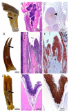
Gill-associated lymphoid tissue (GIALT) of Chondrichthyes and Actinopterygi. All images were taken from the second left gill arch and oriented in the transversal plane of the gill. (AC) Catshark (S. hesperius). (A) Macroscopic image of a dissected gill. Note that the interbranchial septum (S) is continuous with the hypodermis of the outer body surface. The black arrow points to the region as seen in images (B,C). (B) Staining of a transversal sectioned gill arch. Note the thymic tissue with cortex and medulla within the interbranchial septum. (C) Zap-70 staining of a thymic lobe. Note the high numbers of Zap-70+ cells (brown) within the medulla (black arrowhead). (DF) Siberian sturgeon (A. baerii). (D) Macroscopic image of a dissected gill. Note the long but discontinued interbranchial septum (S). The black arrow points to the region as seen in images (E,F). (E) H&E staining of the epithelium lining the distal part of the interbranchial septum. (F) Zap-70 staining of the corresponding region shown in (E). Note the high numbers of Zap-70+ cells (brown) within the epithelium. (GI) Bonefish (Albula vulpes). (G) Macroscopic image of a dissected gill. Note the intermediate length of the interbranchial septum (S). The black arrow points to the region as seen in (H,I). (H) staining of the epithelium lining the distal part of the interbranchial septum. (I) Zap-70 staining of the corresponding region shown in (H). Note the high numbers of Zap-70+ cells (brown) within the epithelium.
Figure 5
Figure 5
Absence of interbranchial lymphoid tissue (ILT) but the presence of gill-associated lymphoid tissue (GIALT) in the interfilamental space in two Neoteleostian species. (AF) European perch (P. fluviatilis). (A) Macroscopic image of gill in transversal projection. The arrowhead points to the end of the interbranchial cleft as also seen in (B,C). Note the short or missing interbranchial septum. (B,C) staining of transversal orientation: no ILT-like structure could be identified in the thin epithelium covering the bottom of the interbranchial cleft. (D) Macroscopic image of gill in a lateral projection. (E,F) Sagittal orientation: Extensive intraepithelial lymphoid aggregates between the proximal part of the filaments at their attachment to the gill arch. The black arrows in (D) show the orientation in the histological sections (E,F), pointing to the transition between the gill arch and the filament where the intraepithelial lymphoid aggregates are situated. (E) H&E staining; black arrowheads point to free lamellae, while white arrowheads point to lamellar structures embedded within the intraepithelial lymphoid aggregate. (F) Immunohistochemical staining using the anti-Zap-70 antibody to indicate Zap-70+ cells (brown). All images were taken from the second left gill arch. (GI) Witch flounder (G. cynoglossus). Sagittal orientation: black arrows show orientation in the histological section pointing to the transition between the gill arch and the filament where intraepithelial lymphoid aggregates are situated. (GH) Extensive intraepithelial lymphoid aggregates between the proximal part of the filament at their attachment to the gill arch. (I) Immunohistochemical staining using the anti-Zap-70 antibody to indicate Zap-70+ cells (brown). All images were taken from a second left gill arch.
Figure 6
Figure 6
Gill-associated lymphoid tissues (GIALT) of zebrafish (D. rerio). Spinning disk confocal deconvolved images of adult zebrafish gills coronal sections across and above the interbranchial septum (A,B’). (A,B’) represent 10 µm maximum intensity projection (MIP) from a multifields stitch (20× objective) displaying phalloidin/DAPI/zap70 labeling for (A,B) and DAPI/zap70 labeling for (A’,B’). Images were acquired from 30 µm thick whole-organism cryosections, while tissues structures are highlighted with phalloidin (F-actin, red) and DAPI (Nuclei, blue) stainings in order to examine the distribution of Zap-70+ cells (white). (A,A’). Zap-70+ cells are mainly concentrated within the afferent aspect of the filaments. Note the high number of Zap-70+ cells within the septum wall, which then assemble into the proximal ILT (pILT) at the uppermost layer of the interbranchial septum. (B,B’). Zap-70+ cells then extend upward from the top of the interbranchial septum to form the distal ILT (dILT) along the afferent aspect of filaments. In addition to the structured lymphoid tissue observed, Zap-70+ cells are also distributed in a diffuse manner among the interlamellar region and the efferent aspect of filaments (A’,B’). F, Filament; La, Lamellae; S, Septum; St, Septum top; Sw, Septum wall; Af, Afferent aspect; Il, interlamellar region; Ef, Efferent aspect; Aa, Afferent artery; Vs, Venous sinus; Ea, Efferent artery; C, Cartilage. Scale bar: 50 µm (A,A’) and 30 µm (B,B’).
Figure 7
Figure 7
Gill-associated lymphoid tissues (GIALT) of medaka (O. latipes). Spinning disk confocal deconvolved images of adult medaka gills paratransversally sectioned (A,A’), coronally sectioned beneath the septum (B,B’), coronally sectioned across the septum (C,C’) and coronally sectioned above the septum (D). (A) is a 10 µm MIP (20× objective). (A’) is a 1 µm MIP (60× objective). (B) is a 1 µm MIP from a 6 fields of view stitch (60× objective) showing two successive gill arches. (B’) is a reoriented and zoomed area from (B). (C) is a 1 µm MIP from a multifields stitch (60× objective). (C’) is a reoriented and zoomed area of (C). (D) is a 1 µm MIP (60× objective). Images were acquired from 30 µm thick whole-organism cryosections, while tissues structures are highlighted with phalloidin (F-actin, red) and DAPI (Nuclei, blue) stainings in order to examine the distribution of Zap-70+ cells (white). (A) Note the almost inexistent interbranchial septum (S). Numerous Zap-70+ cells are seen scattered among gill filaments, lamellae and septa without any structural organization. (A’) Further magnification illustrating the diffuse presence of the Zap-70+ cells within the very short interbranchial septum and both the efferent and the afferent aspects of the filaments. The diffuse distribution of Zap-70+ cells is consistent all along the filaments, with cells present among the afferent aspect, the efferent aspect and the interlamellar region (delimitated by the yellow dotted lines) underneath the septum (B,B’), at the septum (C,C’) and above the septum (D). Ga, Gill arch; F, Filament; La, Lamellae; S, Septum; St, Septum top; Af, Afferent aspect; Il, interlamellar region; Ef, Efferent aspect; Aaa, Afferent arch artery; Aa, Afferent artery; Vs, Venous sinus; Ea, Efferent artery; C, Cartilage; M, skeletal muscles; Sm, Smooth muscle; Pc, Pillar cells; Bl, Blood vessel lumen. Scale bar: 30 µm (AC), 20 µm (A’,D) and 10 µm (B’,C’).
Figure 8
Figure 8
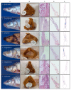
Heads of 6 endemic Labeobarbus species flock in Lake Tana (column (A)) and their isolated gills (column (B)) after 24-h fixation in formalin and followed by 26-year storage in 70% alcohol [24], and a transverse view of lymphoid tissue (ILT) in HE-stained gills (Scale bar is 100 μm, column (C)), transverse view of HE-stained gills (Scale bar is 500 μm, column (D)), transverse drawing of gill morphology (column (E)). The transverse drawings of gill morphology are based on scanned HE-stained gills, and lymphocyte-like cell accumulations are in blue (column (E)).

References

    1. Nelson J.S. Fishes of the World. Wiley; Hoboken, NJ, USA: 2007.
    1. Laurent P. Morphology and physiology of organs of aquatic respiration in vertebrates: The gill. J. Physiol. 1984;79:98–112. - PubMed
    1. Wilson J.M., Laurent P. Fish gill morphology: Inside out. J. Exp. Zool. 2002;293:192–213. doi: 10.1002/jez.10124. - DOI - PubMed
    1. Grevellec A., Tucker A.S. The pharyngeal pouches and clefts: Development, evolution, structure and derivatives. Semin. Cell Dev. Biol. 2010;21:325–332. doi: 10.1016/j.semcdb.2010.01.022. - DOI - PubMed
    1. Kimmel C.B., Ballard W.W., Kimmel S.R., Ullmann B., Schilling T.F. Stages of embryonic development of the zebrafish. Dev. Dyn. 1995;203:253–310. doi: 10.1002/aja.1002030302. - DOI - PubMed