Review
. 2020 Jun 15;21(12):4257.
doi: 10.3390/ijms21124257.

Lipid Mediators Regulate Pulmonary Fibrosis: Potential Mechanisms and Signaling Pathways

Affiliations
Review

Lipid Mediators Regulate Pulmonary Fibrosis: Potential Mechanisms and Signaling Pathways

Vidyani Suryadevara et al. Int J Mol Sci. .

Abstract

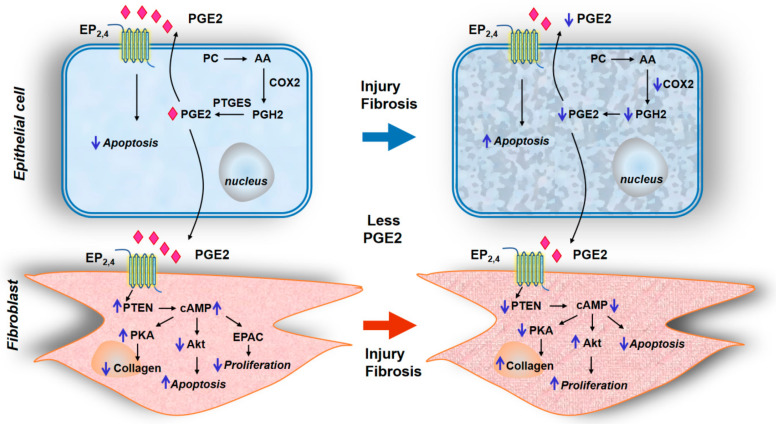
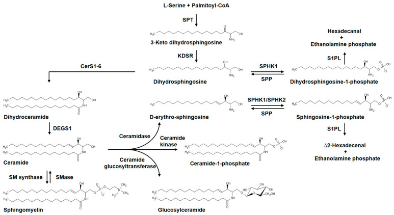
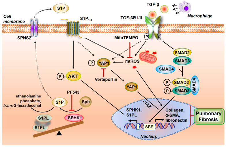
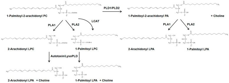
Idiopathic pulmonary fibrosis (IPF) is a progressive lung disease of unknown etiology characterized by distorted distal lung architecture, inflammation, and fibrosis. The molecular mechanisms involved in the pathophysiology of IPF are incompletely defined. Several lung cell types including alveolar epithelial cells, fibroblasts, monocyte-derived macrophages, and endothelial cells have been implicated in the development and progression of fibrosis. Regardless of the cell types involved, changes in gene expression, disrupted glycolysis, and mitochondrial oxidation, dysregulated protein folding, and altered phospholipid and sphingolipid metabolism result in activation of myofibroblast, deposition of extracellular matrix proteins, remodeling of lung architecture and fibrosis. Lipid mediators derived from phospholipids, sphingolipids, and polyunsaturated fatty acids play an important role in the pathogenesis of pulmonary fibrosis and have been described to exhibit pro- and anti-fibrotic effects in IPF and in preclinical animal models of lung fibrosis. This review describes the current understanding of the role and signaling pathways of prostanoids, lysophospholipids, and sphingolipids and their metabolizing enzymes in the development of lung fibrosis. Further, several of the lipid mediators and enzymes involved in their metabolism are therapeutic targets for drug development to treat IPF.

Keywords: G-protein coupled receptors; autotaxin; lipid mediators; lysocardiolipin acyltransferase; lysophosphatidic acid; oxidized phospholipids; phospholipase D; prostaglandins; pulmonary fibrosis; sphingolipids; sphingosine kinase 1; sphingosine-1-phosphate.


Conflict of interest statement

The authors declare no conflict of interest. The funders had no role in the design of the study; in the collection, analyses or interpretation of the data; in the writing of the manuscript or in the decision to publish this review article.

Figures

Figure 1
Figure 1
Prostaglandin E2 signaling via EP2/EP4 in epithelial cells and fibroblasts for the development of pulmonary fibrosis. Schema depicts PGE2 biosynthesis from arachidonic acid by COX2 and prostaglandin E synthases in alveolar epithelial cells. The autacoid function of PGE2 via its receptors EP2 and EP4 regulates homeostatic signaling between the alveolar epithelial cells (AECs) and pulmonary fibroblasts. Fibrotic signaling is specified by enhanced apoptosis and diminished secretion of PGE2 by AECs due to injury, which in turn promotes fibroblast proliferation, collagen deposition, and myofibroblast differentiation, distinct in pulmonary fibrosis. AA—Arachidonic acid, cAMP—cyclic adenosine monophosphate, COX2—Cyclooxygenase-2, EP2,4—Prostaglandin EP2,EP4 receptor, EPAC—Exchange protein directly activated by cAMP, PC—Phosphatidyl choline, PGE2—Prostaglandin E2, PGH2—Prostaglandin H2, PKA—Protein kinase A, PTGES—Prostaglandin E Synthase, PTEN—Phosphatase and tensin homolog.
Figure 2
Figure 2
Sphingolipids implicated in pulmonary fibrosis, their structure, and metabolizing enzymes. An overview of de novo pathways of sphingolipid biosynthesis. Formation of the precursor 3-keto dihydrosphingosine from palmitoyl-CoA and L-serine catalyzed by SPT is the initial rate-limiting step, followed by generation of complex sphingolipids catalyzed by specific metabolic enzymes resulting in the generation of ceramides, sphingosine, sphingosine-1-phosphate (S1P), ceramide-1-phosphate and glucosyl ceramide. S1P or Dihydro S1P is hydrolyzed by S1PL to ∆2-hexadecenal or hexadecanal, respectively, and ethanolamine phosphate. S1P or dihydro S1P is also converted back to sphingosine by SPP. Dysregulation of the metabolic pathway intermediates is involved in the pathogenesis of pulmonary fibrosis. CerS1-6—Ceramide synthase 1-6, DEGS1—Delta 4-Desaturase Sphingolipid 1, KDSR—Keto dihydrosphingosine reductase, SPT—Serine palmitoyltransferase, SMase—Sphingomyelinase, SM synthase—Sphingomyelin synthase, SPHK1—Sphingosine kinase 1, SPHK2—Sphingosine kinase 2, S1P—Sphingosine-1-phosphate, SPP—Sphingosine-1-phosphate phosphatase, S1PL—Sphingosine-1-phosphate lyase.
Figure 3
Figure 3
Crosstalk between S1P and TGF-β signaling cascade in pulmonary fibrosis. Profibrotic signaling by the inflammatory cytokine TGF-β is affected through the dimerization of TGF-β1 and TGF-βII receptors and its phosphorylation and activation of SMADs to induce a fibrogenic transcriptional program. Transport of S1P generated by SPHK1 activation from the cell to the extracellular milieu is enabled by SPNS2 transporter. S1P binding to S1P receptors stimulates mitochondrial reactive oxygen species (mtROS) generation and YAP1 translocation to the nucleus to influence myofibroblast transdifferentiation and matrix remodeling. AKT—Protein kinase B, αSMA—smooth muscle αActin, MitoTEMPO—(2-(2,2,6,6-Tetramethylpiperidin-1-oxyl-4-ylamino) -2-oxoethyl)triphenylphosphonium chloride, PF543—SPHK1 inhibitor, SBE—SMAD-Binding Element, SPNS2—spinster homolog 2, Sph—Sphingosine, SPHK1—Sphingosine kinase 1, S1P—Sphingosine -1-phosphate, S1PL—Sphingosine-1-phosphate lyase, S1P15—Sphingosine-1-phosphate receptors 1–5, SMAD—Mothers Against Decapentaplegic Homolog, TGF-β—Transforming growth factor beta, TGF-β RI/II—Transforming growth factor beta receptor I/II, TAZ—Tafazzin, YAP1—Yes-associated Protein 1.
Figure 4
Figure 4
Generation of lysophosphatidic acid by two different pathways in mammalian cells. Schema showing the conversion of 1-Palmitoyl-2-arachidonyl phosphatidylcholine to LPA by the metabolizing enzymes, PLD, PLA1, PLA2, and autotaxin or lyso PLD. LCAT—lecithin-cholesterol acyltransferase, LPA—lysophosphatidic acid, LPC—lysophosphatidylcholine, lyso PLD—lysophospholipase D, PA—Phosphatidic acid, PC—Phosphatidylcholine, PLA1/2—Phospholipase A1/2, PLD1/2—Phospholipase D 1/2.
Figure 5
Figure 5
Cardiolipin remodeling by lysocardiolipin acyltransferase in pulmonary fibrosis. The mitochondrial phospholipid cardiolipin is converted by iPLA2 or cPLA2 to monolysocardiolipin (MLCL), a regulator of apoptosis, in the mitochondria. Dysregulation of LYCAT function, which remodels the fatty acid composition of cardiolipin, has been implicated in pulmonary fibrosis and Barth syndrome. ALCAT1—Acyl-CoA: lysocardiolipin acyltransferase, CoA—CoenzymeA, FA—Fatty acid, LYCAT—lysocardiolipin acyltransferase, LPC—lysophosphatidylcholine, LPE—lysophosphatidylethanolamine, PC—Phosphatidylcholine, PE—Phosphatidylethanolamine, iPLA2—calcium-independent Phospholipase A2, cPLA2—cytosolic phospholipase A2.
Figure 6
Figure 6
Role of lysophosphatidic signaling via lysophosphatidic acid receptors in pulmonary fibrosis. (A), Alveolar epithelial cell stimulation by lysophosphatidic acid (LPA) activates the G-protein coupled receptor LPA2 and generation of mitochondrial reactive species (mtROS) promotes caspase-3 mediated apoptotic pathway. Rho/ROCK activation by LPA directs the fibrogenic signaling via αvβ6 and TGF-β. (B), In the lung vascular endothelial cells, the LPA/LPA1 activation leads to cytoskeletal changes and VE-cadherin-mediated junctional disruption. (C), LPA/LPA2 signaling in the fibroblasts stimulates the MAPK/ERK and PI3K/AKT pathways, resulting in enhanced TGF-β production and secretion. This results in activation of the TGF-β dependent profibrotic signaling mechanisms via TGF-βI/TGF-βII receptors and activation of SMAD proteins leading to myofibroblast differentiation and collagen production in PF. AKT—Protein kinase B, CNN1—Calponin1, Col1A1—Collagen Type I Alpha 1 chain, ERK—Extracellular signal-regulated kinase, Gαq,12/13,i/o—GTP-binding protein complex alpha subunit, LPA—lysophosphatidic acid, LPA1/2—lysophosphatidic acid receptor type1/2, LPAR—lysophosphatidic acid receptor, mtROS—mitochondrial reactive oxygen species, MAPK—mitogen-activated protein kinase, MLC—myosin light chain, PI3K—Phosphatidylinositol-3-kinase, Ras—RAS family of GTPases, Rho—Rho family of small GTPase, ROCK—Rho-associated coiled-coil containing protein kinase, SMAD2/3/4—Mothers Against Decapentaplegic Homolog 2/3/4, TGF-β—transforming growth factor beta, TGF-β RI/II—transforming growth factor beta receptor I/II, VE—Cadherin vascular endothelial cadherin.

References

    1. Abrass C.K. Cellular lipid metabolism and the role of lipids in progressive renal disease. Am. J. Nephrol. 2004;24:46–53. doi: 10.1159/000075925. - DOI - PubMed
    1. Adibhatla R.M., Hatcher J.F. Role of Lipids in Brain Injury and Diseases. Future Lipidol. 2007;2:403–422. doi: 10.2217/17460875.2.4.403. - DOI - PMC - PubMed
    1. Burnett J.R., Hooper A.J., Hegele R.A. Lipids and cardiovascular disease. Pathology. 2019;51:129–130. doi: 10.1016/j.pathol.2018.12.001. - DOI - PubMed
    1. Sulciner M.L., Gartung A., Gilligan M.M., Serhan C.N., Panigrahy D. Targeting lipid mediators in cancer biology. Cancer Metastasis Rev. 2018;37:557–572. doi: 10.1007/s10555-018-9754-9. - DOI - PubMed
    1. Higenbottam T. Lung lipids and disease. Respiration. 1989;55:14–27. doi: 10.1159/000195747. - DOI - PubMed

LinkOut - more resources