. 2020 Jun 16:20:245.
doi: 10.1186/s12935-020-01326-4. eCollection 2020.

FBXO2 modulates STAT3 signaling to regulate proliferation and tumorigenicity of osteosarcoma cells

Affiliations

FBXO2 modulates STAT3 signaling to regulate proliferation and tumorigenicity of osteosarcoma cells

Xunming Zhao et al. Cancer Cell Int. .

Abstract

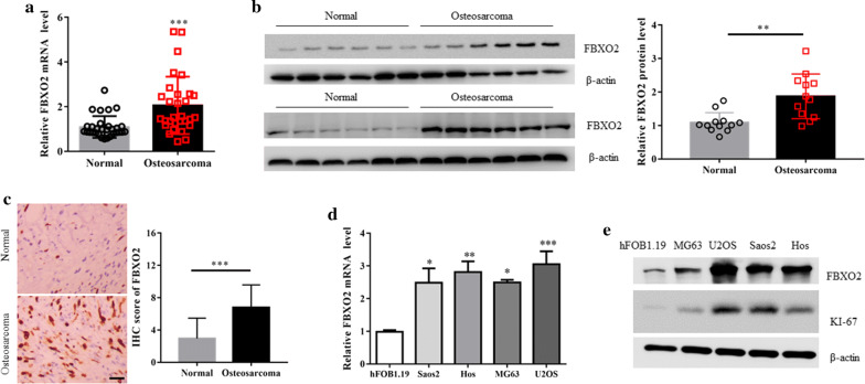
Background: Osteosarcoma (OS) is the most common primary bone malignancy in children and adolescents, and hyperproliferation of cells is a major problem of OS. FBXO2 belongs to the family of F-box proteins, and is a substrate recognition component of the Skp1-Cul1-F-box protein (SCF) E3 ubiquitin ligase complex with specificity for high-mannose glycoproteins. The aim of the present study was to investigate the critical role of FBXO2 in OS cells.

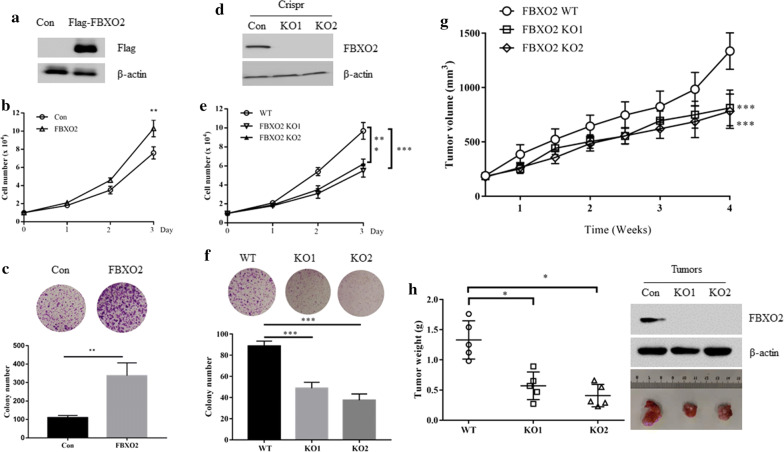
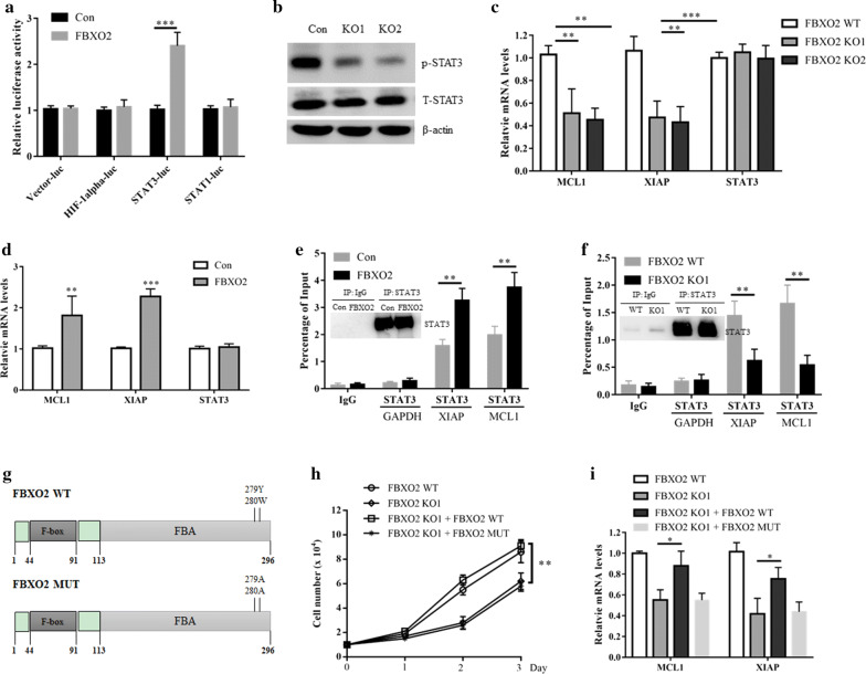
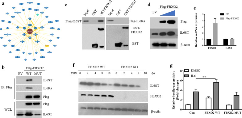
Methods: The protein and mRNA expression levels of FBXO2 in clinic OS patients were measured by quantitative real time-polymerase chain reaction (qRT-PCR), Western blot and Immunohistochemical (IHC) staining assays, respectively. The FBXO2 overexpression model was constructed by retro-virus transfection in OS cells. FBXO2 knockout (KO) cells were generated by Clustered regularly interspaced short palindromic repeat (CRISPR)-CRISPR-associated protein 9 (Cas9) assay. Cell counting and colony formation assays were used to analyze the effect of FBXO2 on the biological function of OS cells. FBXO2 KO cells were injected into nude mice to observe tumor growth in vivo. The interaction between FBXO2 and IL-6 was detected by immunoprecipitation. Luciferase assay was used to determine the transcriptional activity of STAT3.

Results: Here, we show that FBXO2 is significantly up-regulated in clinical OS samples compared to adjacent normal tissues. Ectopic expression of FBXO2 leads to increased OS cell proliferation and colony-forming ability, while FBXO2 knockout by CRISPR-Cas9-based gene editing has the opposite effect. In addition, the glycoprotein recognition activity of FBXO2 is required for its biological function in OS. In vivo experiments showed that FBXO2 knockout greatly impaired the tumorigenicity of OS cells in nude mice. At the molecular level, we found that knocking out FBXO2 can significantly inhibit STAT3 phosphorylation and downstream target gene expression through IL-6R stabilization.

Conclusion: Together, these results indicate that FBXO2 promotes OS development by activating the STAT3 signaling pathway, suggesting that FBXO2 may be a new target for OS treatment.

Keywords: Degradation; FBXO2; IL-6R; Osteosarcoma; STAT3.


Conflict of interest statement

Competing interestsThe authors declare that they have no competing interests.

Figures

Fig. 1
Fig. 1
FBXO2 is upregulated in OS samples and cell lines. a FBXO2 mRNA and b protein expression in OS and adjacent normal tissues. The quantification plot was based on scanning densitometry analysis using the Image J software (v 1.8.0). c Immunohistochemical analyses of 20 OS and adjacent normal tissues using anti-FBXO2 antibody were performed. Representative images of IHC staining were presented. The scale bar represents 50 μm. The quantitative analysis for IHC images were shown. d FBXO2 mRNA and protein e expression in four OS cell lines and normal human osteoblastic hFOB1.19 cells
Fig. 2
Fig. 2
FBXO2 regulates the OS cells proliferation in vitro and in vivo. a FBXO2 protein expression in MG63 cells with or without Flag-FBXO2 overexpression. b Cell growth curve of MG63 cells with or without Flag-FBXO2 overexpression. c Colony formation assay of MG63 cells with or without Flag-FBXO2 overexpression. d FBXO2 KO U2OS cells were generated by CRISPR assay and detected by western blot. e Cell growth curve of FBXO2 WT or FBXO2 KO U2OS cells. f Colony formation assay of FBXO2 WT or FBXO2 KO U2OS cells. g 1 × 107 FBXO2 WT or KO U2OS cells were injected subcutaneously into BALB/c nude mice, tumour growth was measured using a caliper at the indicated times after injection. n = 5 for each group. h Mice were sacrificed 6 weeks after transplantation. The tumors were then excised and weighed and subjected to western blot assay with indicated antibodies
Fig. 3
Fig. 3
FBXO2 activates STAT3 signaling in OS dependent on its glycoprotein recognizing activity. a U2OS cells were transfected with the indicated plasmids for 36 h and then cells were lysed for luciferase assay. The results are indicated as fold induction of luciferase activity from triplicate experiments. ***p < 0.001. b U2OS cells were transfected with FBXO2 plasmid for 36 h, cells were lysed for western blot analysis. c The mRNA levels of MCL1, XIAP and STAT3 in FBXO2 WT or KO U2OS cells. d The mRNA levels of MCL1, XIAP and STAT3 in U2OS cells transfected with or without FBXO2. e ChIP assay showed that overexpression of FBXO2 increased the binding of STAT3 to the promotors of both MCL1 and XIAP genes in U2OS cells. The enrichment efficiency of endogenous STAT3 protein was detected by western blot assay. f ChIP assay showed that in FBXO2 KO U2OS cells, the binding of STAT3 to the promotors of both MCL1 and XIAP genes was decreased. The enrichment efficiency of endogenous STAT3 protein was detected by western blot. g The diagram of FBXO2 WT and FBXO2 MUT. FBXO2 WT or FBXO2 MUT plasmids were transfected into U2OS FBXO2 KO cells for 36 h. h The cell growth curve and i the mRNA levels of MCL1 and XIAP in these cells
Fig. 4
Fig. 4
FBXO2 interacts with interleukin 6 receptor to stabilize it. a The interaction protein network of FBXO2 revealed by the BioGRID database. b 293T cell were transfected with EV, Flag-FBXO2 WT or MUT for 36 h. Cells were lysed and immunoprecipitated with Flag M2 beads. The immunoprecipitates were detected by western blot using indicated antibodies. c Beads coated with bacterially expressed GST or GST-SENP2 were incubated with purified Flag-IL6ST or Flag-IL6Ra protein. Beads were washed, and the bound proteins were analyzed by Western blotting with indicated antibodies. d Immunoblotting analysis of the cell lysates of MG63 cells transfected with increased doses of FBXO2WT. e qRT-PCR analysis of IL6ST and FBXO2 in MG63 cells transfected with EV or Flag-FBXO2 WT. f Cell lysates from FBXO2 WT and KO U2OS cells treated with 20 μg/ml cycloheximide (CHX) were subjected to western blot with the indicated antibodies. g U2OS cells were co-transfected STAT3-Luc with indicated plasmids. 24 h after transfection, cells were treated with DMSO or IL-6 (5 ng/ml) for additional 12 h. The cells were lysed and assayed for luciferase assay as above described. **p < 0.01

References

    1. Jackson TM, Bittman M, Granowetter L. Pediatric malignant bone tumors: a review and update on current challenges, and emerging drug targets. Curr Probl Pediatr Adolesc Health Care. 2016;46(7):213–228. doi: 10.1016/j.cppeds.2016.04.002. - DOI - PubMed
    1. Ballatori SE, Hinds PW. Osteosarcoma: prognosis plateau warrants retinoblastoma pathway targeted therapy. Signal Transduct Target Ther. 2016;1:16001. doi: 10.1038/sigtrans.2016.1. - DOI - PMC - PubMed
    1. Botter SM, Neri D, Fuchs B. Recent advances in osteosarcoma. Curr Opin Pharmacol. 2014;16:15–23. doi: 10.1016/j.coph.2014.02.002. - DOI - PubMed
    1. Hattinger CM, Fanelli M, Tavanti E, Vella S, Ferrari S, Picci P, Serra M. Advances in emerging drugs for osteosarcoma. Expert Opin Emerg Drugs. 2015;20(3):495–514. doi: 10.1517/14728214.2015.1051965. - DOI - PubMed
    1. Ciechanover A. The unravelling of the ubiquitin system. Nat Rev Mol Cell Biol. 2015;16(5):322–324. doi: 10.1038/nrm3982. - DOI - PubMed