Vascular depression for radiology: A review of the construct, methodology, and diagnosis
- PMID: 32549954
- PMCID: PMC7288775
- DOI: 10.4329/wjr.v12.i5.48
Vascular depression for radiology: A review of the construct, methodology, and diagnosis
Abstract
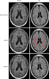
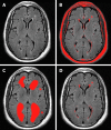
Vascular depression (VD) as defined by magnetic resonance imaging (MRI) has been proposed as a unique subtype of late-life depression. The VD hypothesis posits that cerebrovascular disease, as characterized by the presence of MRI-defined white matter hyperintensities, contributes to and increases the risk for depression in older adults. VD is also accompanied by cognitive impairment and poor antidepressant treatment response. The VD diagnosis relies on MRI findings and yet this clinical entity is largely unfamiliar to neuroradiologists and is rarely, if ever, discussed in radiology journals. The primary purpose of this review is to introduce the MRI-defined VD construct to the neuroradiology community. Case reports are highlighted in order to illustrate the profile of VD in terms of radiological, clinical, and neuropsychological findings. A secondary purpose is to elucidate and elaborate on the measurement of cerebrovascular disease through visual rating scales and semi- and fully-automated volumetric methods. These methods are crucial for determining whether lesion burden or lesion severity is the dominant pathological contributor to VD. Additionally, these rating methods have implications for the growing field of computer assisted diagnosis. Since VD has been found to have a profile that is distinct from other types of late-life depression, neuroradiologists, in conjunction with psychiatrists and psychologists, should consider VD in diagnosis and treatment planning.
Keywords: Case reports; Cerebrovascular disorders; Depression; Magnetic resonance imaging; Neuroradiology; Vascular depression; White matter hyperintensities.
©The Author(s) 2020. Published by Baishideng Publishing Group Inc. All rights reserved.
Conflict of interest statement
Conflict-of-interest statement: All authors deny any conflicts of interest related to this work.
Figures


References
-
- Alexopoulos GS, Meyers BS, Young RC, Campbell S, Silbersweig D, Charlson M. 'Vascular depression' hypothesis. Arch Gen Psychiatry. 1997;54:915–922. - PubMed
-
- Steffens DC, Krishnan KR. Structural neuroimaging and mood disorders: recent findings, implications for classification, and future directions. Biol Psychiatry. 1998;43:705–712. - PubMed
-
- Hickie I, Scott E, Mitchell P, Wilhelm K, Austin MP, Bennett B. Subcortical hyperintensities on magnetic resonance imaging: clinical correlates and prognostic significance in patients with severe depression. Biol Psychiatry. 1995;37:151–160. - PubMed
-
- Krishnan KR, McDonald WM. Arteriosclerotic depression. Med Hypotheses. 1995;44:111–115. - PubMed
-
- Krishnan KR, Hays JC, Blazer DG. MRI-defined vascular depression. Am J Psychiatry. 1997;154:497–501. - PubMed
Publication types
LinkOut - more resources
Full Text Sources

