

Can Ellipsoid Sign be One of the Earliest Findings of the Medial Femoral Condyle Chondromalacia of Knee Antero Posterior X-Ray?
- PMID: 32549968
- PMCID: PMC7270317
- DOI: 10.1007/s43465-020-00140-4
Can Ellipsoid Sign be One of the Earliest Findings of the Medial Femoral Condyle Chondromalacia of Knee Antero Posterior X-Ray?
Abstract
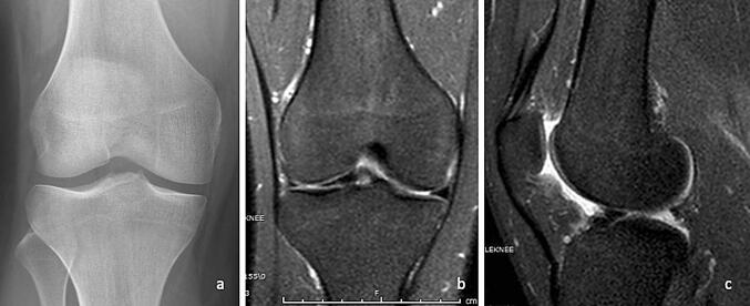
Background/purpose of the study: Aim of the study was to investigate whether ellipsoid sign (ES) in the region corresponding to the cartilaginous facet of the medial femoral condyle (MFC) indicates early cartilage lesion. The finding has not been defined in the literature yet.
Methods: The study was performed retrospectively with 50 patients who underwent articular cartilage examination and arthroscopy between 2015 and 2018. Patients were divided into two according to the presence or absence of ES. There were 24 patients in case group (Group A) and 26 patients in controls (Group B). Weight-bearing cartilage areas of MFC in the region where ES was found in both groups were classified according to arthroscopic Outerbridge classification (OC) and compared statistically with each other.
Results: There was no significant difference in terms of age, gender and alignment between Group A and Group B (p > 0.05). All OC grades were higher in Group A (p < 0.001). Positive correlation between ES and MFC chondromalacia grade was detected (r: 0.671, t: 6.266, p < 0.05).
Conclusion: ES, which refers to the difference in bone density in MFC seen in antero-posterior (AP) X-Ray, is a result of changes in subchondral bone due to chronic cartilage loss. ES detected on AP X-Ray may help in early diagnosis of medial femoral articular cartilage chondromalacia, even at grade 0 or 1. We recommend searching for ES, as the earliest symptom of chondromalacia, which occurs even before MRI lesions. Future studies may reveal additional information about ES.
Keywords: Cartilage; Chondromalacia; Ellipsoid sign; Femoral condyle; Osteoarthritis.
© Indian Orthopaedics Association 2020.
Conflict of interest statement
Conflict of interestAll authors approved the manuscript and all authors declare that are no conflicts of interest regarding the publication.
Figures


Similar articles
-
Topographic Matching of Osteochondral Allograft Transplantation Using Lateral Femoral Condyle for the Treatment of Medial Femoral Condyle Lesions: A Computer-Simulated Model Study.Arthroscopy. 2018 Nov;34(11):3033-3042. doi: 10.1016/j.arthro.2018.05.039. Arthroscopy. 2018. PMID: 30392687
-
Quantitative Magnetic Resonance Imaging UTE-T2* Mapping of Cartilage and Meniscus Healing After Anatomic Anterior Cruciate Ligament Reconstruction.Am J Sports Med. 2014 Aug;42(8):1847-56. doi: 10.1177/0363546514532227. Epub 2014 May 8. Am J Sports Med. 2014. PMID: 24812196 Free PMC article.
-
Topographic Analysis of Lateral Versus Medial Femoral Condyle Donor Sites for Oblong Medial Femoral Condyle Lesions.Arthroscopy. 2020 Nov;36(11):2900-2908. doi: 10.1016/j.arthro.2020.07.007. Epub 2020 Jul 28. Arthroscopy. 2020. PMID: 32735941
-
Articular cartilage lesions in the symptomatic anterior cruciate ligament-deficient knee.Arthroscopy. 2003 Sep;19(7):685-90. doi: 10.1016/s0749-8063(03)00403-1. Arthroscopy. 2003. PMID: 12966374 Review.
-
Arthroscopic evaluation of articular cartilage lesions in posterior-cruciate-ligament-deficient knees.Arthroscopy. 2003 Mar;19(3):262-8. doi: 10.1053/jars.2003.50037. Arthroscopy. 2003. PMID: 12627150 Review.
References
-
- Disler G, McCauley TR, Kelman. CG, Fuschs MD, Ratner LM, Wirth CR, et al. Fat suppressed three dimensional spoiled gradient-echo MR imaging of hyaline cartilage defects in the knee: Comparison with Standart MR imaging and arthroscopy. AJR Am J Roentgenol 1996;167:127–132. - PubMed
-
- McCaulay RT, Recht MP, Disler DG. Clinical imaging of articular cartilage in the knee. Sem Musc Skel Rad. 2001;5:293–304. - PubMed
-
- Cicuttini F, Forbes A, Asbeutah A, Morris K, Stuckey S. Comprasion and reproducibility of fast and conventional spoiled gradient-echo magnetic resonance sequences in the determination of knee cartilage volume. Journal of Orthopaedic Research. 2000;18:580–584. doi: 10.1002/jor.1100180410. - DOI - PubMed
LinkOut - more resources
Full Text Sources
Research Materials