Review
. 2020 Jun 9:6:36.
doi: 10.1038/s41421-020-0167-x. eCollection 2020.

Inflammasome activation and regulation: toward a better understanding of complex mechanisms

Affiliations
Review

Inflammasome activation and regulation: toward a better understanding of complex mechanisms

Danping Zheng et al. Cell Discov. .

Abstract

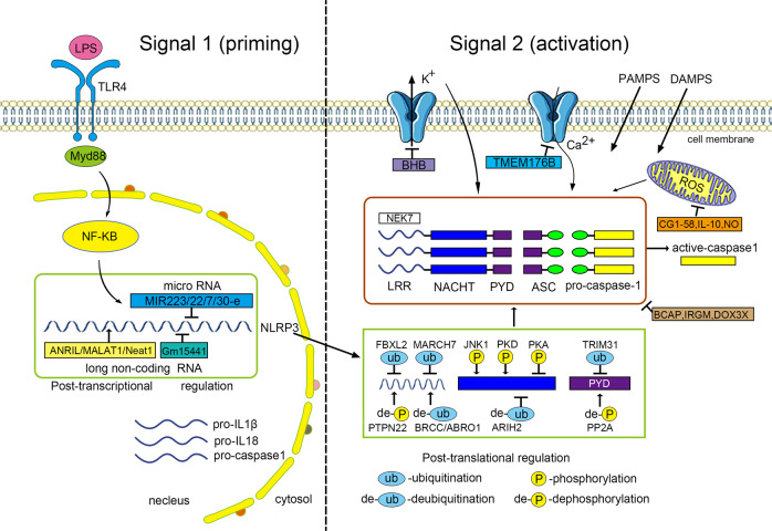
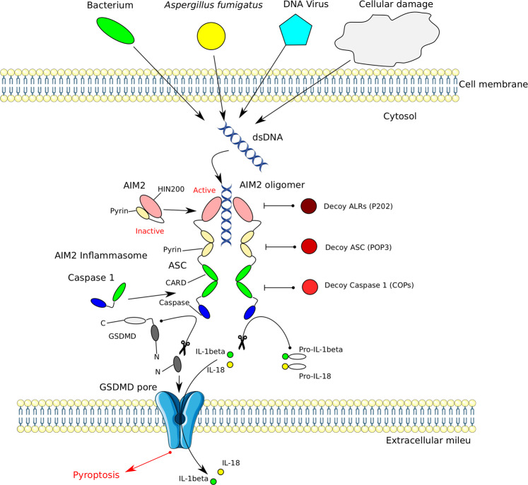
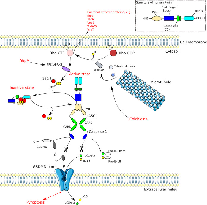
Inflammasomes are cytoplasmic multiprotein complexes comprising a sensor protein, inflammatory caspases, and in some but not all cases an adapter protein connecting the two. They can be activated by a repertoire of endogenous and exogenous stimuli, leading to enzymatic activation of canonical caspase-1, noncanonical caspase-11 (or the equivalent caspase-4 and caspase-5 in humans) or caspase-8, resulting in secretion of IL-1β and IL-18, as well as apoptotic and pyroptotic cell death. Appropriate inflammasome activation is vital for the host to cope with foreign pathogens or tissue damage, while aberrant inflammasome activation can cause uncontrolled tissue responses that may contribute to various diseases, including autoinflammatory disorders, cardiometabolic diseases, cancer and neurodegenerative diseases. Therefore, it is imperative to maintain a fine balance between inflammasome activation and inhibition, which requires a fine-tuned regulation of inflammasome assembly and effector function. Recently, a growing body of studies have been focusing on delineating the structural and molecular mechanisms underlying the regulation of inflammasome signaling. In the present review, we summarize the most recent advances and remaining challenges in understanding the ordered inflammasome assembly and activation upon sensing of diverse stimuli, as well as the tight regulations of these processes. Furthermore, we review recent progress and challenges in translating inflammasome research into therapeutic tools, aimed at modifying inflammasome-regulated human diseases.

Keywords: Cell signalling; NOD-like receptors.


Conflict of interest statement

Conflict of interestThe authors declare that they have no conflict of interest.

Figures

Fig. 1
Fig. 1. Pattern diagram for inflammasome assembly and activation.
Inflammasomes can be activated by a multitude of infectious and sterile stimuli, including microbiome-derived signals (e.g., bacteria, fungi, parasites, and viruses) and host-derived signals (e.g., ion flux, mitochondrial dysfunction, ROS, and metabolic factors). Upon activation, the inflammasome sensors NAIP/NLRC4, NLRP3/6/7, AIM2/IHI16 initiate the canonical inflammasome assembly by recruiting and forming pro-caspase-1 filaments, with or without the ASC adapter. The assembly of non-canonical inflammasomes involves the pro-caspase-11 (−4/5 in humans) or pro-caspase-8. Consequently, the active caspase-1 or caspase-8 leads to the maturation and secretion of inflammatory cytokines IL-1β and IL-18. The active capsase-1 or caspase-11/4/5 triggers the cleavage of GSDMD, which can either cause pyroptosis or activate the NLRP3 inflammasome complex. In addition, the active caspase-8 mediates another effector function, apoptosis.
Fig. 2
Fig. 2. Regulation of the NLRP3 inflammasome activation.
Activation of the NLRP3 inflammasome involves both the priming and activation steps. In the priming step (Signal 1), inflammatory triggers such as TLR4 agonist LPS induces the NF-κB-mediated expression of NLRP3, pro-IL-1β and pro-caspase-1. In the activation step, diverse PAMPs and DAMPs trigger the NLRP3 inflammasome assembly though unifying events such as ion flux, mitochondrial dysfunction and ROS formation. Regulation of the NLRP3 inflammasome activation can occur at the post-transcriptional and post-translational levels. MicroRNAs (e.g., MiR-223, MiR-22, MiR-7, and MiR-30e) inhibit the NLRP3 activity by targeting its UTR binding sites, and long non-coding RNAs (e.g., ANRIL, MALAT1, Neat 1, and Gm15441) can either promote or inhibit the inflammasome signaling. The post-translational modifications at different sites and domains of NLRP3 protein include phosphorylation, dephosphorylation, ubiquitination, de-ubiquitination, and S-nitrosylation. Many molecules such as BCAP, IRGM, and DDX3X can interfere the assembly of NLRP3, ASC, and procaspase-1. Other negative regulatory molecules inhibit the inflammasome activation by targeting the K+ efflux (e.g., BHB), cytosolic Ca2+ (e.g., TMEM176B), mitochondrial function (e.g., macrophage CGI-58, IL-10, and NO).
Fig. 3
Fig. 3. Activation and regulation of the AIM2 inflammasome.
AIM2 is composed of the N-terminal pyrin (PYD) and the C-terminal HIN200 domains. Interaction of both domains renders the molecule inactive. Binding of free cytosolic double-stranded (ds)DNA releases auto-inhibition with subsequent oligomerization, recruitment of adapter ASC via PYD–PYD interaction and polymerization of the ASC protein. The dsDNA instigating the cascade can originate from microorganisms of different kingdoms and from host cellular damage. Through ASC polymerization pro-caspase-1 is recruited to the complex via CARD–CARD interactions. This induces the maturation and secretion of IL-1β and IL-18, and results in pyroptosis. AIM2 inflammasome signaling can be inhibited on several levels by decoy proteins. Decoy ALRs such as P202, which lack a PYD, may interfere with AIM2 binding to dsDNA and may also inhibit AIM2 oligomerization. POPs are decoy ASC proteins inhibit PYD–PYD interactions between ASC and AIM2. Decoy caspase-1 (COPs) lack a caspase domain and inhibit caspase-1 recruitment to the inflammasome by blocking CARD–CARD association.
Fig. 4
Fig. 4. Structure, activation, and regulation of the Pyrin inflammasome.
Pyrin is kept autoinhibited by 14-3-3 proteins which are bound to the phosphorylated pyrin. The inactivating pyrin phosphorylation is maintained by the kinases PRK1 and PRK2. The kinases are activated by RhoA GTPase. Inactivation of RhoA by bacterial effector proteins leads to dephosphorylation of PRK1/2. Subsequently, pyrin is de-phosphorylated, leading to dissociation of 14-3-3 and pyrin activation. Activated pyrin recruits ASC via PYD–PYD interactions. The complex then associates with pro-caspase-1 through CARD-CARD interactions. Active caspase-1 mediates IL-1β and IL-18 maturation, as well as pyroptosis. Yersinia outer protein M (YopM) suppresses the pyrin inflammasome activation by employing PRK1/2. Microtubule polymerization inhibition by colchicine leads to release of GEF-H1 which renders RhoA GTPase active, resulting in inactivating phosphorylation of pyrin by PRK1/2.

References

    1. Medzhitov R. Origin and physiological roles of inflammation. Nature. 2008;454:428–435. doi: 10.1038/nature07201. - DOI - PubMed
    1. Janeway CA, Jr, Medzhitov R. Innate immune recognition. Annu Rev. Immunol. 2002;20:197–216. doi: 10.1146/annurev.immunol.20.083001.084359. - DOI - PubMed
    1. Schroder K, Tschopp J. The inflammasomes. Cell. 2010;140:821–832. doi: 10.1016/j.cell.2010.01.040. - DOI - PubMed
    1. Akira S, Takeda K. Toll-like receptor signalling. Nat. Rev. Immunol. 2004;4:499–511. doi: 10.1038/nri1391. - DOI - PubMed
    1. Geijtenbeek TB, Gringhuis SI. Signalling through C-type lectin receptors: shaping immune responses. Nat. Rev. Immunol. 2009;9:465–479. doi: 10.1038/nri2569. - DOI - PMC - PubMed