Review
. 2020 Jun 5:5:11.
doi: 10.1038/s41536-020-0096-1. eCollection 2020.

Innervation: the missing link for biofabricated tissues and organs

Affiliations
Review

Innervation: the missing link for biofabricated tissues and organs

Suradip Das et al. NPJ Regen Med. .

Abstract

Innervation plays a pivotal role as a driver of tissue and organ development as well as a means for their functional control and modulation. Therefore, innervation should be carefully considered throughout the process of biofabrication of engineered tissues and organs. Unfortunately, innervation has generally been overlooked in most non-neural tissue engineering applications, in part due to the intrinsic complexity of building organs containing heterogeneous native cell types and structures. To achieve proper innervation of engineered tissues and organs, specific host axon populations typically need to be precisely driven to appropriate location(s) within the construct, often over long distances. As such, neural tissue engineering and/or axon guidance strategies should be a necessary adjunct to most organogenesis endeavors across multiple tissue and organ systems. To address this challenge, our team is actively building axon-based "living scaffolds" that may physically wire in during organ development in bioreactors and/or serve as a substrate to effectively drive targeted long-distance growth and integration of host axons after implantation. This article reviews the neuroanatomy and the role of innervation in the functional regulation of cardiac, skeletal, and smooth muscle tissue and highlights potential strategies to promote innervation of biofabricated engineered muscles, as well as the use of "living scaffolds" in this endeavor for both in vitro and in vivo applications. We assert that innervation should be included as a necessary component for tissue and organ biofabrication, and that strategies to orchestrate host axonal integration are advantageous to ensure proper function, tolerance, assimilation, and bio-regulation with the recipient post-implant.

Keywords: Regenerative medicine; Tissue engineering.


Conflict of interest statement

Competing interestsD.K.C and H.C.L. are co-founders of Axonova Medical, LLC, and D.K.C. is a co-founder of INNERVACE, Inc., which are University of Pennsylvania spin-out companies focused on translation of advanced regenerative therapies to treat nervous system disorders. Multiple patents relate to the composition, methods, and use of the constructs described in the paper, including U.S. Patent 9,895,399 (D.K.C.), U.S. Patent App. 15/032,677 (D.K.C.), U.S Provisional Patent App. 62/322,434 (D.K.C., H.I.C., M.D.S.), U.S. Provisional Patent 62/569,255 (D.K.C), and U.S. Provisional Patent App. 62/758,203 (D.K.C, S.D., W.G.V.). No other author declares competing interests.

Figures

Fig. 1
Fig. 1. Innervation of cardiac, skeletal, and smooth muscle.
The table (left) presents a summary of innervation in the development and functional regulation of the heart (cardiac muscle), GI tract and bladder (smooth muscle), and skeletal muscle, and tissue engineering approaches for these tissues and cases where innervation has been considered in these strategies. The schematic (right) presents parasympathetic and sympathetic inputs to the organs surveyed in this manuscript; only connections with one side are shown for simplicity. We also show somatic fibers connected with skeletal muscle (bottom right). Preganglionic parasympathetic fibers emanate mostly from the brainstem, except for innervation to the lower large intestine and the bladder, which originate from the sacral spinal cord. Most preganglionic parasympathetic nerves synapse with ganglia in close proximity to the target organ (shown as blue circles near or at the organs). On the other hand, preganglionic sympathetic fibers exit from the gray matter in the thoracic or lumbar spinal cord and interact with ganglia in the bilateral sympathetic chain (yellow). In other cases, preganglionic fibers within the splanchnic nerves pass through the sympathetic chain to synapse further on with abdominal ganglia. CN cranial nerve, CG celiac ganglia, SMG superior mesenteric ganglion, IMG inferior mesenteric ganglion.
Fig. 2
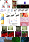
Fig. 2. Strategies to promote innervation of biofabricated constructs.
ae Innervation of skeletal muscle grafts by neurotization. a Schematic of the repair of an abdominal wall defect in mice with a PLLA/PLGA porous scaffold seeded with myoblasts, endothelial cells, and fibroblasts. Neurotized grafts were innervated by suturing to the graft the proximal femoral bundle including the nerve. b, c Images of the neurotized and control grafts, respectively, with the white arrowheads indicating the grafts and the white arrows marking the femoral nerve. d Assessment of compound muscle action potential (CMAP) amplitude in the grafts after electrical stimulation of the attached femoral bundle. e Neuromuscular junctions in the grafts were stained for acetylcholine receptors (AChR; red) and synapses (synaptophysin; green). (Reprinted with permission from Kaufman and Kaplan et al.; Copyright John Wiley & Sons). f, g Diagrams of examples of traditional strategies for promoting peripheral nerve regeneration using nerve guidance conduits modified to deliver neurotrophic/growth factors or cells using biomaterial-based delivery methods. (Reprinted with permission from Daly et al.; Copyright The Royal Society). hm Nerve guidance conduit functionalized for drug release to promote nerve regeneration. h Schematic of a nerve guidance conduit sutured to the proximal and distal ends of a damaged nerve to promote axon regrowth. i Halloysite nanotube (HNTs)-based conduits were loaded with 4-aminopyridine (4-AP), a potassium channel blocker that promotes neurotransmitter release and extends action potentials. j Conduits composed of drug-loaded chitosan, epichlorohydrin-crosslinked chitosan, chitosan/HNT composites, or crosslinked composites were analyzed for in vitro 4-AP release. Data presented as mean ± standard error of the mean. k Image of the repair of a rat sciatic nerve defect with a conduit. l, m Hematoxylin and eosin stain image of a longitudinal and cross-sectional view of the conduit, respectively, 4 weeks post repair showing regenerating nerve with infiltrating Schwann cells (conduit material in dark red). (Reprinted with permission from Manoukian et al.; Copyright Elsevier). nq Neurotrophic factor overexpression for the reinnervation of injured myocardium. Rat hearts denervated by cryoinjury were injected with adenoviruses encoding n, o GFP or p, q GDNF. After 5 days, the hearts were n, p whole-mount imaged for neurofilament-M (NFM) or o, q immunolabeled to denote cardiomyocytes (α-actinin (AA); red), GDNF (blue), and axons (NFM; green). Hearts with GFP overexpression showed only sparse axon presence, while GDNF overexpression led to a significant presence of axons in the injured area and axon growth into the myocardium. (Reprinted with permission from Miwa et al.; open access, PLOS). rt Engineered scaffolds with smooth muscle sheets co-cultured with neural progenitor cells for GI repair. r Image of the scaffold 14 days after subcutaneous implantation in the back of athymic rats. The scaffold consisted of human smooth muscle cells cultured on molds to promote alignment and human enteric neuronal progenitor cells added on top of the muscle sheets. The sheets were wrapped around a tubular scaffold of chitosan and collagen to resemble the structure of the gut. The engineered neuromuscular tissue was stained to show s contractile smooth muscle (caldesmon; green) and t differentiated neurons (β-tubulin III; green). (Reprinted with permission from Zakhem et al.; Copyright Elsevier). Scale bars: e 10 µm; l, m, s, t 100 µm; n, p 1 mm; o, q 50 µm.
Fig. 3
Fig. 3. Stretch-grown nerve grafts and their application for the directed innervation of biofabricated constructs.
a Stretch-grown tissue-engineered nerve grafts (TENGs) are fabricated employing a mechanobioreactor that has a towing membrane and a mobile towing block. b Aggregates of neurons are seeded on two sides of the towing membrane, allowed to grow connecting axons, and then separated at a specific rate by pulling the towing block to stretch the axons. c, d Phase contrast images of TENGs before and after application of mechanical forces to stretch the axon tracts. e, f Confocal images of aggregates and axon tracts, respectively, stained for nuclei (Hoechst; blue), axons (β-tubulin III; green), motor neurons (p75; red). (Reprinted with permission from Katiyar et al.; authors of content, John Wiley & Sons). g TENGs are embedded in collagen and encased in a nerve conduit before implantation in a nerve injury model. (Reprinted with permission from Huang and Cullen et al.; authors of content, Mary Ann Liebert, Inc.). h, i After implantation to repair a peripheral nerve lesion, host axons (SMI31; purple) were seen extending along the path dictated by TENG axons (GFP; green), thus demonstrating the mechanism of axon-facilitated axon regeneration. (Reprinted with permission from Struzyna et al.; open access, Wolters Kluwer Medknow Publications). j Distal outgrowth of implanted TENG axons (GFP+) into the host nerve was observed 6 weeks post repair. Bundles of host axons (NF-200; red) projected along the construct and also into the distal nerve (yellow arrow). k, l Higher magnification images showing axonal bundles and ganglia in the implanted TENGs, respectively. (Reprinted with permission from Huang and Cullen et al.; authors of content, Mary Ann Liebert, Inc.). m Stretch-grown axons may be incorporated into tissue-specific biofabricated constructs by first obtaining the main cell types and neurons associated with each type of tissue. Abbreviations: superior cervical ganglia (SCG), celiac ganglia (CG), submandibular ganglia (SMdG). n (1) After creating a scaffold seeded with tissue-specific cells, (2) neurons can be mechanically aggregated by centrifugation, plated on the towing membrane and cultured to ensure attachment of their neurites to cells in the scaffold. (3) Axons projected from the aggregates would be stretch-grown by controlling the rate at which the towing membrane is pulled away, thus creating a construct with directed innervation that could be used for in vitro or in vivo applications. Scale bars: c, d 1000 µm; e, f 500 µm; h 25 µm; i 6 µm; j 100 µm; k, l 25 µm.
Fig. 4
Fig. 4. Application of micro-tissue engineered neural networks (micro-TENNs) for peripheral reinnervation.
a, b Phase contrast images of a unidirectional and bidirectional micro-TENN at 5 days, respectively, consisting of an aggregate of rat embryonic cortical neurons seeded at one (unidirectional) or both (bidirectional) ends of a hydrogel micro-column. These aggregates extend aligned axonal tracts throughout the extracellular matrix-filled lumen. c Confocal image of a micro-TENN with neurons from the cerebral cortex at 28 days and stained for axons (β-tubulin III; red), somata/dendrites (MAP2; green), and nuclei (Hoechst; blue). d, e High magnification images of the aggregate and axon tract regions of the micro-TENN construct, respectively. (Reprinted with permission from Serruya et al.; authors of content, John Wiley and Sons). f Bidirectional micro-TENN at 14 days fabricated with an aggregate of rat embryonic ventral midbrain neurons (left) and striatal neurons (right) and stained for dopaminergic neurons (tyrosine hydroxylase (TH); red), medium spiny (striatal) neurons (DARPP-32; green), synapses (synapsin I; purple), and nuclei (Hoechst; blue). gi Zoom-ins show the dopaminergic aggregate, outgrowth from medium spiny neurons, and physical integration of dopaminergic axons with the striatal target, respectively. (Reprinted with permission from Struzyna et al.; authors of content, Copyright John Wiley and Sons). j Micro-TENNs may serve to promote regeneration of axonal connections from spinal motor neurons to muscles suffering volumetric muscle loss (VML). In this application, micro-TENNs can be microinjected at the proximal nerve and with engineered muscle distally to guide axon growth from the nerve back to the muscle belly to regenerate lost neuromuscular connections. k Micro-TENNs could be sourced from autonomic ganglia to serve as parallel pathways to native autonomic innervation that could project axons to innervate the target organs. Scale bars: a, b 100 µm; c 200 µm; d, e 100 µm; f 250 µm; g 50 µm.

Similar articles

Cited by

References

    1. Mai, J. K. & Paxinos, G. The Human Nervous System (Academic Press, 2011).
    1. Cardinali, D. P. Autonomic Nervous System: Basic and Clinical Aspects (Springer, 2017).
    1. Mulroney, S. & Myers, A. Netter’s Essential Physiology E-Book (Elsevier Health Sciences, 2015).
    1. Furness JB, Callaghan BP, Rivera LR, Cho H-J. The enteric nervous system and gastrointestinal innervation: integrated local and central control. Adv. Exp. Med. Biol. 2014;817:39–71. - PubMed
    1. Kreipke RE, Birren SJ. Innervating sympathetic neurons regulate heart size and the timing of cardiomyocyte cell cycle withdrawal. J. Physiol. 2015;593:5057–5073. - PMC - PubMed