Review
. 2020 Jun 8:8:23.
doi: 10.1038/s41413-020-0099-y. eCollection 2020.

Molecular mechanosensors in osteocytes

Affiliations
Review

Molecular mechanosensors in osteocytes

Lei Qin et al. Bone Res. .

Abstract

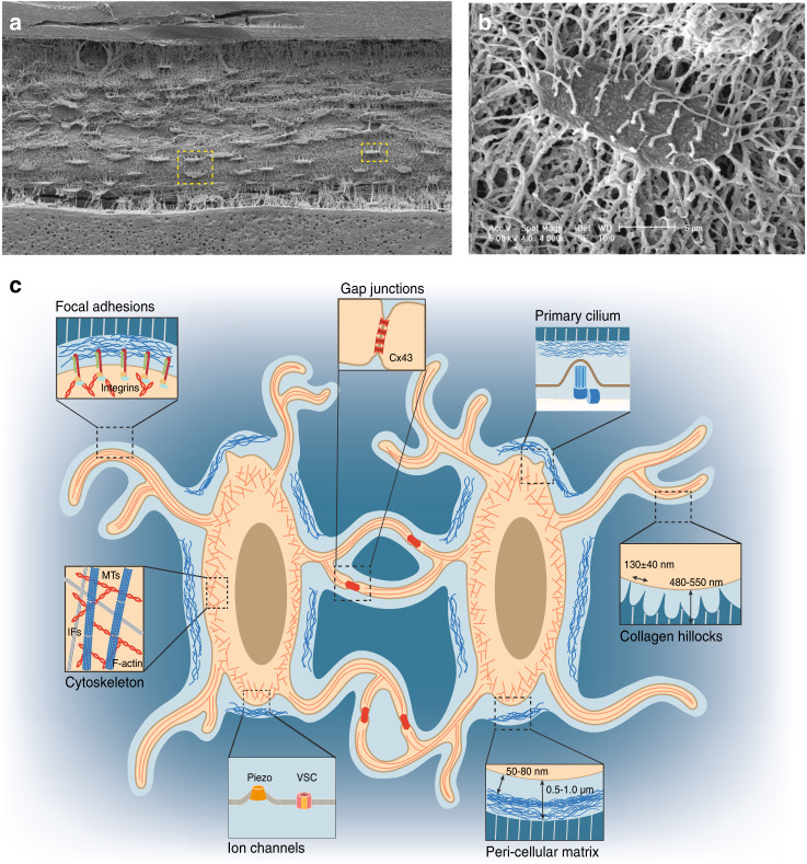
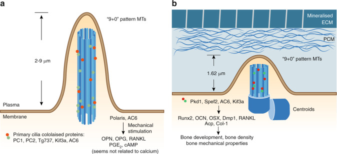
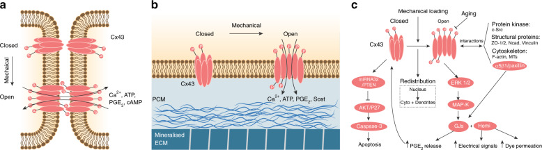
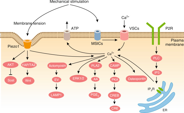
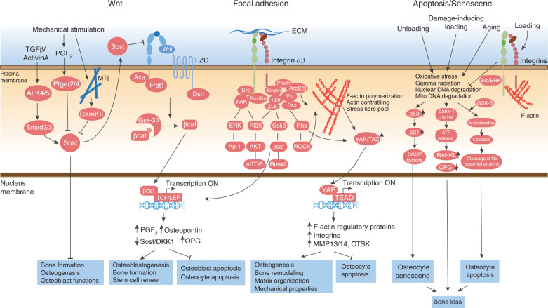
Osteocytes, the most abundant and long-lived cells in bone, are the master regulators of bone remodeling. In addition to their functions in endocrine regulation and calcium and phosphate metabolism, osteocytes are the major responsive cells in force adaptation due to mechanical stimulation. Mechanically induced bone formation and adaptation, disuse-induced bone loss and skeletal fragility are mediated by osteocytes, which sense local mechanical cues and respond to these cues in both direct and indirect ways. The mechanotransduction process in osteocytes is a complex but exquisite regulatory process between cells and their environment, between neighboring cells, and between different functional mechanosensors in individual cells. Over the past two decades, great efforts have focused on finding various mechanosensors in osteocytes that transmit extracellular mechanical signals into osteocytes and regulate responsive gene expression. The osteocyte cytoskeleton, dendritic processes, Integrin-based focal adhesions, connexin-based intercellular junctions, primary cilium, ion channels, and extracellular matrix are the major mechanosensors in osteocytes reported so far with evidence from both in vitro and in vitro studies. This review aims to give a systematic introduction to osteocyte mechanobiology, provide details of osteocyte mechanosensors, and discuss the roles of osteocyte mechanosensitive signaling pathways in the regulation of bone homeostasis.

Keywords: Bone quality and biomechanics; Osteoporosis.


Conflict of interest statement

Competing interestsThe authors declare no competing interests.

Figures

Fig. 1
Fig. 1
Osteocytes in the LCS of the bone environment. a SEM image of acid-etched resin-embedded cortical bone sections reveals an ellipsoid cell shape and extensive canaliculi connections among osteocytes.b Magnified SEM image of a single osteocyte highlighted in the yellow square in a. c Illustration of osteocytes in the LCS of the bone environment. Magnified cartoon image of two adjacent osteocytes highlighted in the yellow square in a. The important aspects of osteocytes are highlighted in magnified cartoon images: focal adhesions, gap junctions, the primary cilium, cell cytoskeleton, ion channels, pericellular matrix at the lacunar region, and collagen hillocks at the canalicular region. [Panels a and b from Bonewald et al., reprinted with permission]
Fig. 2
Fig. 2
In vivo models commonly used in osteocyte mechanobiology. Examples of active loading models with the right ulna (a) and right tibia (b) in mice. Loading model mice were under avertin-induced anesthesia, and the right sides of the ulna and tibia were subjected to cyclic mechanical compression with a computationally controlled machine. The contralateral left limbs served as controls. Illustrations of ulna and tibia loading are highlighted in yellow and green boxes, respectively. c Illustration of the HLU model in mice. Experimental mice were outfitted with a tail harness, and their hindlimbs were suspended within customized cages. [panel c from Robling et al., reprinted with permission]
Fig. 3
Fig. 3
Illustration of cytoskeleton and Integrin subunits in a single osteocyte. Three types of cytoskeletal components are observed in osteocytes: IFs are mainly reported in the cell body, and F-actin and MTs are reported in both the cell body and dendrites. Compared with total MTs, detyrosinated MTs seem to be more localized to osteocyte processes and the primary cilium. The focal adhesion proteins Integrins show distinct distributions in osteocytes: Integrin β1 is mainly localized to the plasma membrane of the cell body, whereas Integrin β3 is primarily localized to the surface of dendrites
Fig. 4
Fig. 4
Focal adhesions on the osteocyte cell body and dendrites. a Heterodimers of Integrin β3 with Integrins α1/2/3/4/5 are localized to the osteocyte cell body surface. The heads of these heterodimers contact the pericellular matrix, and their tails are linked to the F-actin cytoskeleton. Moreover, the classical focal adhesion components Vinculin and Paxillin were reported to colocalize with Integrin β1-mediated focal adhesions.b Heterodimers of Integrin β3 with Integrin αv are localized to osteocyte dendritic surfaces. The purinergic channel pannexin 1, the ATP-gated purinergic receptor P2X7R and the low-voltage transiently opened T-type calcium channel CaV3.2-1 reside in close proximity to Integrin β3 attachment foci.c Illustration of major focal adhesion components at cell-ECM interphases.,
Fig. 5
Fig. 5
The osteocyte primary cilium in mechanobiology. a Illustration of the primary cilia from in vitro cultured osteocyte-like cells. The primary cilium is a unique cell protrusion structure consisting of nine doublet microtubules in the form of a “9 + 0” pattern., In cultured MLOY4 cells, this cilia-like structure was shown to be 2–9 μm in length., Several ciliary proteins, such as PC1, PC2, Tg737, and Kif3a, colocalize in this structure. Among them, Polaris and AC6 were reported to participate in osteocyte responses to mechanical stimulation.b Illustration of the primary cilium in vivo from the embedded osteocytes of bone sections. Unlike the results of in vitro detection, in vivo recordings of the primary cilium showed a morphological change of the cell membrane in which the mother centriole contacts the plasma membrane and a very short axoneme forms a cilium-like protrusion. With Aα-Tub staining and confocal imaging, primary cilia in osteocytes were measured and found to have an average length of 1.62 μm. The ciliary proteins Pkd1, Spef2, AC6, and Kif3a also participate in osteocyte mechanical bone adaptation
Fig. 6
Fig. 6
Osteocyte gap junctions and hemichannels in mechanobiology. a Illustration of osteocyte GJs in response to mechanical stimulation. A hexameric array of six connexin subunits gives rise to a connexon, and two juxtaposed connexons on the surfaces of adjacent cells form a GJ., When osteocytes experience mechanical stimulation, the Cx43 protein is phosphorylated, and the connexon is opened, allowing the exchange of several effectors, such as calcium, ATP, PGE2, and cAMP, between connecting cells. b Illustration of osteocyte hemichannels in response to mechanical stimulation. Unopposed connexons called hemichannels at the cell membrane act as direct conduits between the cytosol and extracellular environment.c Signaling pathways involved in Cx43-based GJs and hemichannels during osteocyte mechanobiology
Fig. 7
Fig. 7
Illustration of ion channels involved in osteocyte mechanobiology. During osteocyte mechanotransduction, the earliest event that takes place is an increase in the intracellular Ca2+ concentration of the cells. This calcium mobilization process is first triggered by the activation of MSICs. Among all the MSICs, Piezo1 is a promising mechanogating ion channel in osteocyte mechanobiology. Piezo1 is a curved channel that is highly engaged with the cell membrane. Mechanical stimulation increases the osteocyte membrane tension, which further induces the opening of Piezo1 channels. Downstream effectors of Piezo1 channels include the Akt–Sost pathway, YAP/TAZ–Wnt pathway, and intracellular calcium signaling. Upon MSIC opening, ions are exchanged between the cytoplasm and extracellular environment. This process further changes the plasma membrane charge balance and induces the opening of VSCs. Interestingly, the calcium that undergoes flux induced by mechanical stimulation is derived from not only external fluid and medium but also sites of internal calcium storage, such as the endoplasmic reticulum. This calcium mobilization can activate downstream effectors, such as actomyosin, Erk1/2, PGE2, PAK, and osteopontin. Calcium mobilization also regulates ATP release in osteocytes upon mechanical stimulation.
Fig. 8
Fig. 8
Signaling pathways involved in osteocyte mechanobiology. The Wnt/β-Catenin pathway mechanistically, the canonical Wnt/β-Catenin pathway is activated through the binding of Wnt ligands to a coreceptor complex consisting of Lrp5 or Lrp6 and FZD., This binding further activates the intercellular effector Dsh by FZD-mediated phosphorylation. Activated Dsh leads to the phosphorylation of Gsk-3β, which inhibits free β-Catenin in the cytosol by phosphorylating β-Catenin at multiple serine/threonine sites. Once Gsk-3β is phosphorylated by Dsh, it releases captured β-Catenin. As a result, free β-Catenin is translocated to nuclei, where it binds the coeffectors Tcf and Lef, inducing downstream gene transcription. Downstream effects of β-Catenin include the expression of Wnt target genes and secretory proteins (Opg, Osteopontin), and load-induced PGE2 secretion. Sclerostin antagonizes Wnt signaling through its competitive binding to Lrp5 and Lrp6 at their first two YWTD-EGF repeat domains. Mechanical stimulation can suppress Sost expression through both Peger2/4 and the MT pathway. In addition, the Tgfβ-Smad2/3 pathway can enhance sclerostin expression. As a result, during the osteocyte mechanotransduction process, the Wnt/β-Catenin pathway enhances osteoblastogenesis and bone formation; however, sclerostin negatively regulates the Wnt/β-Catenin pathway. Focal adhesion As the central proteins in the FA complex, Integrins, especially Integrin β subunits, are essential for bone development and osteocyte mechanotransduction. The “Integrin adhesome” is a network of 156 proteins in the FA complex. In the FA complex, Kindlin-2, Talin, and other structural proteins are directly linked to the cytoplasmic tail of the Integrin β subunit, which further connects with the Pinch, Paxillin, Vinculin, and Arp2/3 proteins., This Integrin adhesome complex links the ECM and F-actin cytoskeleton and enhances the activation of downstream pathways, such as the Erk, PI3K, Gsk3, and Rho pathways. Upon F-actin cytoskeleton polymerization, YAP/TAZ coordinate signals from Rho GTPase and tension of the actomyosin cytoskeleton, initiate downstream target gene expression, and finally enhance osteogenesis and bone remodeling and inhibit osteocyte apoptosis. Apoptosis/senescence osteocyte apoptosis, a form of programmed cell death, and senescence, a death-resistant cell fate program, are common features of aging bone tissue. Appropriate mechanical stimulation prevents osteocyte apoptosis, whereas aging, damage-inducing loading and disuse induce osteocyte apoptosis and senescence through several different pathways. In contrast, mechanical stimulation induces Src/Erk activation through Integrin and the cytoskeleton in osteocytes, inhibits apoptotic and senescence-related pathways and supports osteocyte survival.

References

    1. Bonewald LF. The amazing osteocyte. J. Bone Min. Res. 2011;26:229–238. doi: 10.1002/jbmr.320. - DOI - PMC - PubMed
    1. Manolagas SC. Birth and death of bone cells: basic regulatory mechanisms and implications for the pathogenesis and treatment of osteoporosis. Endocr. Rev. 2000;21:115–137. - PubMed
    1. Schaffler MB, Kennedy OD. Osteocyte signaling in bone. Curr. Osteoporos. Rep. 2012;10:118–125. doi: 10.1007/s11914-012-0105-4. - DOI - PMC - PubMed
    1. Schaffler MB, Cheung WY, Majeska R, Kennedy O. Osteocytes: master orchestrators of bone. Calcif. Tissue Int. 2014;94:5–24. doi: 10.1007/s00223-013-9790-y. - DOI - PMC - PubMed
    1. Florencio-Silva R, Sasso GR, Sasso-Cerri E, Simoes MJ, Cerri PS. Biology of bone tissue: structure, function, and factors that influence bone cells. Biomed. Res. Int. 2015;2015:421746. doi: 10.1155/2015/421746. - DOI - PMC - PubMed