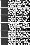
Dense-UNet: a novel multiphoton in vivo cellular image segmentation model based on a convolutional neural network
- PMID: 32550136
- PMCID: PMC7276369
- DOI: 10.21037/qims-19-1090
Dense-UNet: a novel multiphoton in vivo cellular image segmentation model based on a convolutional neural network
Abstract
Background: Multiphoton microscopy (MPM) offers a feasible approach for the biopsy in clinical medicine, but it has not been used in clinical applications due to the lack of efficient image processing methods, especially the automatic segmentation technology. Segmentation technology is still one of the most challenging assignments of the MPM imaging technique.
Methods: The MPM imaging segmentation model based on deep learning is one of the most effective methods to address this problem. In this paper, the practicability of using a convolutional neural network (CNN) model to segment the MPM image of skin cells in vivo was explored. A set of MPM in vivo skin cells images with a resolution of 128×128 was successfully segmented under the Python environment with TensorFlow. A novel deep-learning segmentation model named Dense-UNet was proposed. The Dense-UNet, which is based on U-net structure, employed the dense concatenation to deepen the depth of the network architecture and achieve feature reuse. This model included four expansion modules (each module consisted of four down-sampling layers) to extract features.
Results: Sixty training images were taken from the dorsal forearm using a femtosecond Ti:Sa laser running at 735 nm. The resolution of the images is 128×128 pixels. Experimental results confirmed that the accuracy of Dense-UNet (92.54%) was higher than that of U-Net (88.59%), with a significantly lower loss value of 0.1681. The 90.60% Dice coefficient value of Dense-UNet outperformed U-Net by 11.07%. The F1-Score of Dense-UNet, U-Net, and Seg-Net was 93.35%, 90.02%, and 85.04%, respectively.
Conclusions: The deepened down-sampling path improved the ability of the model to capture cellular fined-detailed boundary features, while the symmetrical up-sampling path provided a more accurate location based on the test result. These results were the first time that the segmentation of MPM in vivo images had been adopted by introducing a deep CNN to bridge this gap in Dense-UNet technology. Dense-UNet has reached ultramodern performance for MPM images, especially for in vivo images with low resolution. This implementation supplies an automatic segmentation model based on deep learning for high-precision segmentation of MPM images in vivo.
Keywords: Skin in vivo; U-Net; dense-UNet; image segmentation; multiphoton microscopy (MPM).
2020 Quantitative Imaging in Medicine and Surgery. All rights reserved.
Conflict of interest statement
Conflicts of Interest: All authors have completed the ICMJE uniform disclosure form (available at http://dx.doi.org/10.21037/qims-19-1090). HL reports grants received during the conduct of the study from the Canadian Institutes of Health Research, grants from the Canadian Dermatology Foundation, the VGH & UBC Hospital Foundation, and the BC Hydro Employees Community Services Fund; also, HL has a patent with Zeng H, Lui H, McLean D, Lee A, Wang H, and Tang S: Apparatus and Methods for Multiphoton Microscopy. US patent # 9687152B2, June 27, 2017 Canadian Patent # 2832162; issued May 14, 2019. HZ reports grants received during the conduct of the study from the Canadian Institutes of Health Research, the Canadian Dermatology Foundation, the VGH & UBC Hospital Foundation, and from the BC Hydro Employees Community Services Fund; also, HZ has a patent with Zeng H, Lui H, McLean D, Lee A, Wang H, and Tang S: Apparatus and Methods for Multiphoton Microscopy. US patent # 9687152B2, June 27, 2017 Canadian Patent # 2832162; issued May 14, 2019. YW reports grants received during the conduct of the study from the National Natural Science Foundation of China. GC reports grants received outside the submitted work from the United Fujian Provincial Health and Education Project for Tackling the Key Research of China, the Natural Science Foundation of Fujian Province, the Program for Changjiang Scholars and Innovative Research Team in University, the Special Funds of the Central Government Guiding Local Science and Technology Development, and the Scientific Research Innovation Team Construction Program of Fujian Normal University. The other authors have no conflicts of interest to declare.
Figures


References
LinkOut - more resources
Full Text Sources
Miscellaneous
