

Late diagnosis of generalized lymphangiomatosis in a woman presenting with respiratory distress
- PMID: 32550956
- PMCID: PMC7292890
- DOI: 10.1016/j.radcr.2020.05.021
Late diagnosis of generalized lymphangiomatosis in a woman presenting with respiratory distress
Abstract
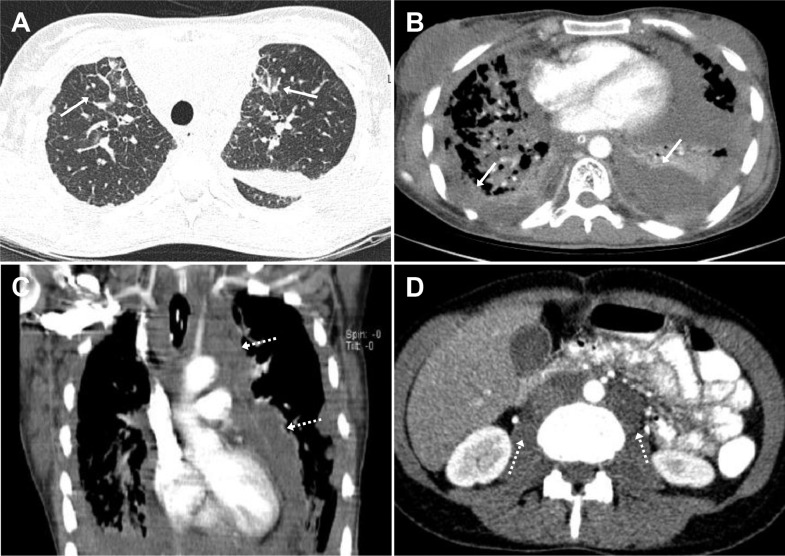
Generalized lymphangiomatosis (GLA) is a rare lymphatic abnormality, mostly affects children and young individuals and can be a diagnostic challenge because of wide spectrum of clinical manifestations. A 26-year-old woman presented to the emergency department of our institution with respiratory distress and hypoxia. The patient reported similar episodes for the past 10 years without a definite diagnosis. The imaging study demonstrated findings suggestive of GLA with pulmonary, retroperitoneal and osseous involvements which was confirmed on pathological studies from a lung biopsy. A concise review of the clinical, imaging and pathological findings of GLA is provided in this study. A comprehensive history and physical examination, laboratory and pathological work up and imaging is required to make the diagnosis of GLA. The characteristic imaging findings play an essential role to rule out other possible diagnoses and raise the possibility of GLA.
Keywords: Chylothorax; Generalized Lymphangiomatosis; Lymphangioma; Lymphatic abnormality; Pulmonary lymphangiectasis.
© 2020 The Authors. Published by Elsevier Inc. on behalf of University of Washington.
Figures


References
-
- Faber D.L., Galili R., Nitzan O., Sharoni E. Systemic generalized lymphangiomatosis: a diagnostic challenge. Isr Med Assoc J. 2015;17:785–786. - PubMed
-
- Arda K.N., Akay S., Kizilkanat K.T. Generalized cystic lymphangiomatosis incidentally recognized in an asymptomatic adult: peroperative, CT, MRI, and histopathological findings of a very rare case. Niger J Clin Pract. 2019;22:1778–1780. - PubMed
Publication types
LinkOut - more resources
Full Text Sources
Miscellaneous
