

Visual recovery and vascular reperfusion after vaso-occlusive retinopathy from anti-phospholipid syndrome associated with systemic lupus erythematosus
- PMID: 32551401
- PMCID: PMC7287235
- DOI: 10.1016/j.ajoc.2020.100763
Visual recovery and vascular reperfusion after vaso-occlusive retinopathy from anti-phospholipid syndrome associated with systemic lupus erythematosus
Abstract
Purpose: To report a case of visual recovery and vascular reperfusion after vaso-occlusive retinopathy from anti-phospholipid syndrome associated with systemic lupus erythematosus.
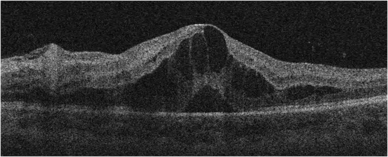
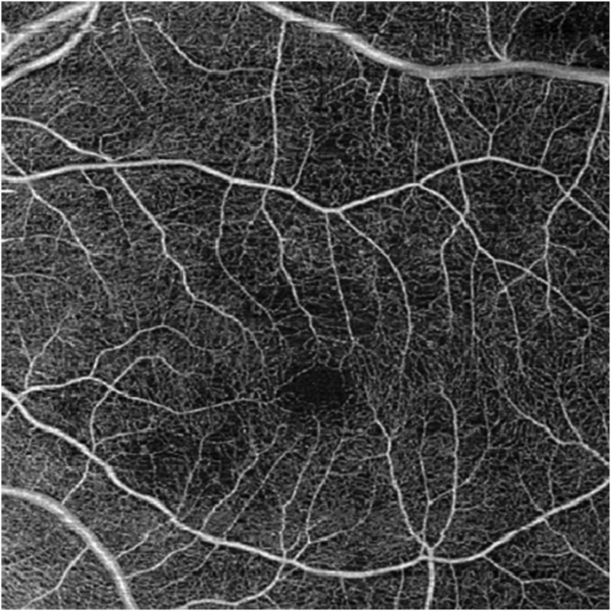
Observations: A 15-year-old boy with a known diagnosis of systemic lupus erythematosus and a clinically significant anti-phospholipid panel presented with sudden vision loss in the left eye. Examination and ocular imaging revealed signs of vaso-occlusive retinopathy. The patient was immediately started on high dose intravenous steroids, followed by mycophenolate mofetil. He remained on aspirin. After showing no improvement in retinal arteriole and capillary perfusion he was started on therapeutic anti-coagulation with enoxaparin. He regained 20/20 vision. Intravenous fluorescein angiography demonstrated reperfusion of retinal arterioles. Optical coherence tomography angiography showed return of flow in the capillary networks.
Conclusions: We present a case of vaso-occlusive retinopathy in a patient with known systemic lupus erythematosus and a clinically significant anti-phospholipid panel, thus meeting criteria for anti-phospholipid syndrome. He was treated with intravenous methylprednisolone, mycophenolate motefil, aspirin, and enoxaparin. The patient not only had great recovery of visual acuity, but also demonstrated reperfusion of arterioles and reconstitution of flow in the retinal capillary network. These findings suggest that the vaso-occlusive disease is reversible if the diagnosis is made promptly and intensive therapy is initiated.
Importance: Currently there are no reported cases of vaso-occlusive retinopathy from APLS and SLE with visual recovery, reperfusion, and return of capillary flow.
Keywords: APLS; Case report; Systemic lupus erythematosus; Vaso-occlusive retinopathy.
© 2020 The Authors.
Figures


References
-
- Silpa-archa S., Lee J.J., Foster C.S. Ocular manifestations in systemic lupus erythematosus. Br J Ophthalmol. 2016;100(1):135–141. - PubMed
-
- Peponis V., Kyttaris V.C., Tyradellis C., Vergados I., Sitaras N.M. Ocular manifestations of systemic lupus erythematosus: a clinical review. Lupus. 2006;15(1):3–12. - PubMed
-
- Acharya N., Pineda R., Uy H.S., Foster C.S. Discoid lupus erythematosus masquerading as chronic blepharoconjunctivitis. Ophthalmology. 2005;112(5):e19–23. - PubMed
-
- Miyakis S., Lockshin M.D., Atsumi T. International consensus statement on an update of the classification criteria for definite antiphospholipid syndrome (APS) J Thromb Haemost JTH. 2006;4(2):295–306. - PubMed
-
- Taraborelli M., Leuenberger L., Lazzaroni M.G. The contribution of antiphospholipid antibodies to organ damage in systemic lupus erythematosus. Lupus. 2016;25(12):1365–1368. - PubMed
