Review
. 2020 Aug 1;319(2):L277-L288.
doi: 10.1152/ajplung.00195.2020. Epub 2020 Jun 17.

Novel insights on the pulmonary vascular consequences of COVID-19

Affiliations
Review

Novel insights on the pulmonary vascular consequences of COVID-19

François Potus et al. Am J Physiol Lung Cell Mol Physiol. .

Abstract

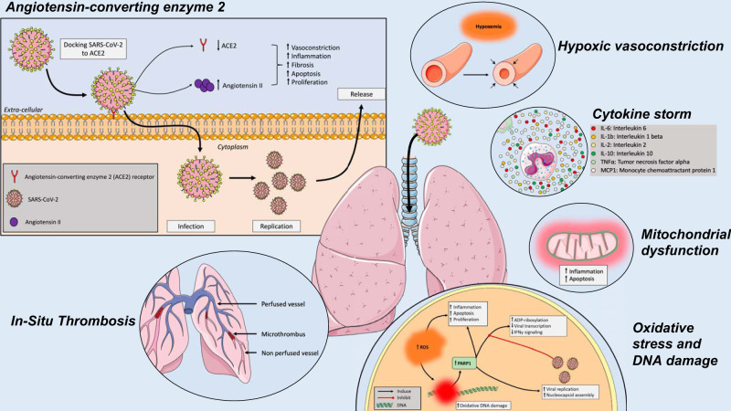
In the last few months, the number of cases of a new coronavirus-related disease (COVID-19) rose exponentially, reaching the status of a pandemic. Interestingly, early imaging studies documented that pulmonary vascular thickening was specifically associated with COVID-19 pneumonia, implying a potential tropism of the virus for the pulmonary vasculature. Moreover, SARS-CoV-2 infection is associated with inflammation, hypoxia, oxidative stress, mitochondrial dysfunction, DNA damage, and lung coagulopathy promoting endothelial dysfunction and microthrombosis. These features are strikingly similar to what is seen in pulmonary vascular diseases. Although the consequences of COVID-19 on the pulmonary circulation remain to be explored, several viruses have been previously thought to be involved in the development of pulmonary vascular diseases. Patients with preexisting pulmonary vascular diseases also appear at increased risk of morbidity and mortality. The present article reviews the molecular factors shared by coronavirus infection and pulmonary vasculature defects, and the clinical relevance of pulmonary vascular alterations in the context of COVID-19.

Keywords: COVID-19; SARS-CoV-1; SARS-CoV-2; coronavirus; pulmonary vascular diseases; vascular remodeling.


Conflict of interest statement

S.P. reports grants from Actelion, grants from AstraZeneca, grants from Resverlogix, outside of the submitted work. M.L. reports personal fees from Air Liquide Health Care, outside the submitted work. S.P. received speaker fees from Actelion Pharmaceuticals and unrestricted grant from Actelion Pharmaceuticals, AstraZeneca (in-kind), Glaxo-Smith-Kline and Resverlogix outside the context of the submitted work. M.L. received speaker fees, and nonfinancial support from Air Liquide Healthcare and SEFAM outside the context of the submitted work. None of the other authors has any conflicts of interest, financial or otherwise, to disclose.

Figures

Fig. 1.
Fig. 1.
Effects of the severe acute respiratory syndrome coronavirus 2 (SARS-CoV-2) on the lungs and the pulmonary vasculature. SARS-CoV-2 cycle starts with the interaction with the angiotensin-converting enzyme 2 (ACE2) receptor from the host cells. Upon entry into the host cells, the viral RNA genome is then translated and proteins and RNA are packaged into progeny virions being released to infect more cells. The SARS-CoV-2-related disease (COVID-19) results in cytokine outburst, including IL-6, IL-1b, IL-2, IL-10, TNF-α, and monocyte chemoattractant protein-1 (MCP-1). Reactive oxygen species (ROS) are key signaling molecules that play an important role in the progression of inflammatory disorders, resulting in oxidative stress, mitochondrial dysfunction, and DNA damage. In addition to parenchymal abnormalities, disseminated intravascular coagulation, endothelial dysfunction, and impaired hypoxic pulmonary vasoconstriction ultimately generate pulmonary microthrombi, ventilation-perfusion mismatch, and hypoxemia.
Fig. 2.
Fig. 2.
Effects of the severe acute respiratory syndrome coronavirus 2 (SARS-CoV-2) on pulmonary hemodynamics and the heart. Acute cardiac injury, manifesting as an ejection fraction decline, and troponin and natriuretic peptide elevation are commonly observed in patients admitted with SARS-CoV-2-related disease (COVID-19). Diastolic dysfunction has also been described in the acute stage of SARS-CoV-1 in patients without preexisting cardiac disease. Whether cardiac injury in COVID-19 results from direct myocardial infection, stress cardiomyopathy, high demand in patients with significantly increased afterload due to secondary pulmonary hypertension in the context of hypoxemia and limited myocardial reserve, or indirect myocardial injury due to secondary hemophagocytic lymphohistiocytosis and microvascular alterations requires further evaluation.

Similar articles

Cited by

References

    1. Ackermann M, Verleden SE, Kuehnel M, Haverich A, Welte T, Laenger F, Vanstapel A, Werlein C, Stark H, Tzankov A, Li WW, Li VW, Mentzer SJ, Jonigk D. Pulmonary vascular endothelialitis, thrombosis, and angiogenesis in COVID-19. N Engl J Med. In press. doi:10.1056/NEJMoa2015432. - DOI - PMC - PubMed
    1. Ahn DG, Shin HJ, Kim MH, Lee S, Kim HS, Myoung J, Kim BT, Kim SJ. Current status of epidemiology, diagnosis, therapeutics, and vaccines for novel coronavirus disease 2019 (COVID-19). J Microbiol Biotechnol 30: 313–324, 2020. doi:10.4014/jmb.2003.03011. - DOI - PMC - PubMed
    1. Ai T, Yang Z, Hou H, Zhan C, Chen C, Lv W, Tao Q, Sun Z, Xia L. Correlation of chest CT and RT-PCR testing in coronavirus disease 2019 (COVID-19) in China: a report of 1,014 cases. Radiology. In press. doi:10.1148/radiol.2020200642. - DOI - PMC - PubMed
    1. Alexander PE, Debono VB, Mammen MJ, Iorio A, Aryal K, Deng D, Brocard E, Alhazzani W. COVID-19 coronavirus research has overall low methodological quality thus far: case in point for chloroquine/hydroxychloroquine. J Clin Epidemiol 123: 120–126, 2020. doi:10.1016/j.jclinepi.2020.04.016. - DOI - PMC - PubMed
    1. Alhogbani T. Acute myocarditis associated with novel Middle east respiratory syndrome coronavirus. Ann Saudi Med 36: 78–80, 2016. doi:10.5144/0256-4947.2016.78. - DOI - PMC - PubMed

MeSH terms