. 2020 Oct;63(4):478-489.
doi: 10.1165/rcmb.2020-0107OC.

ATF4 Mediates Mitochondrial Unfolded Protein Response in Alveolar Epithelial Cells

Affiliations

ATF4 Mediates Mitochondrial Unfolded Protein Response in Alveolar Epithelial Cells

Dingyuan Jiang et al. Am J Respir Cell Mol Biol. 2020 Oct.

Abstract

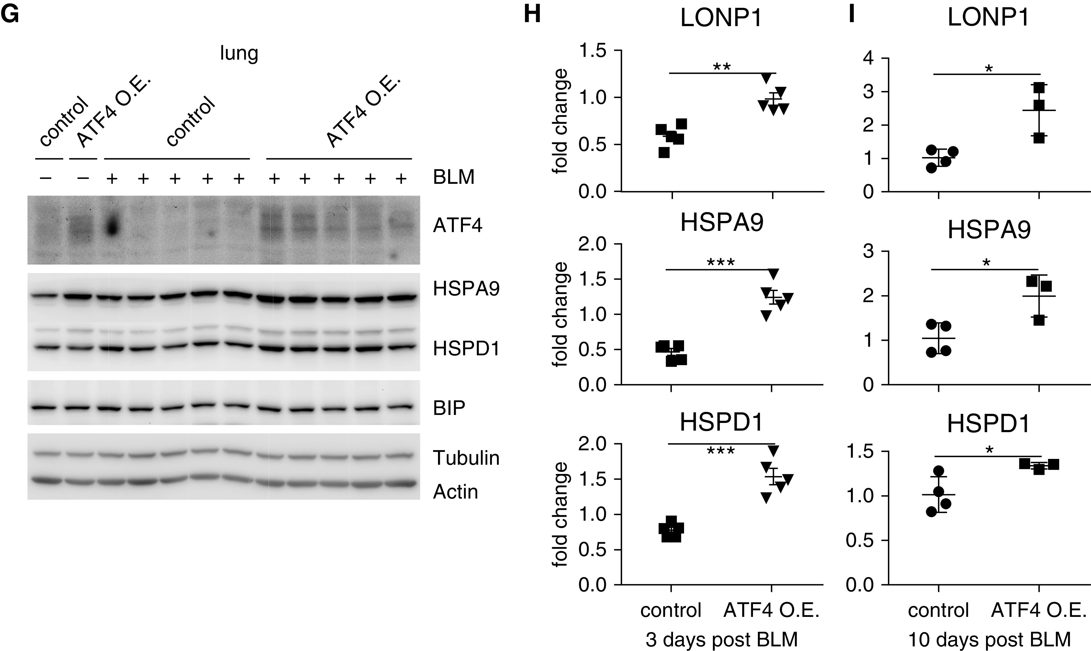
Although endoplasmic reticulum (ER) unfolded protein response (UPRER) is well known, mitochondrial unfolded protein response (UPRmt) has not been recognized in alveolar epithelial cells. Furthermore, ER stress and mitochondrial dysfunction are frequently encountered in alveolar epithelial cells from an array of lung disorders. However, these two scenarios have been often regarded as separate mechanisms contributing to the pathogeneses. It is unclear whether there is interplay between these two phenomena or an integrator that couples these two signaling cascades in the stressed alveolar epithelial cells from those pathologies. In this study, we defined UPRmt in alveolar epithelial cells and identified ATF4 (activating transcription factor 4), but not ATF5, as the key regulator of UPRmt. We found that UPRER led to UPRmt and mitochondrial dysfunction in an ATF4-dependent manner. In contrast, mitochondrial stresses did not activate UPRER. We found that alveolar epithelial ATF4 and UPRmt were induced in aged mice with experimental pulmonary fibrosis as well as in patients with idiopathic pulmonary fibrosis. Finally, we found that the inducible expression of ATF4 in mouse alveolar epithelial cells aggravated pulmonary UPRmt, lung inflammation, body weight loss, and death upon bleomycin-induced lung injury. In conclusion, ER stress induces ATF4-dependent UPRmt and mitochondrial dysfunction, indicating a novel mechanism by which ER stress contributes to the pathogeneses of a variety of pulmonary disorders.

Keywords: ER stress; alveolar epithelial cell; mitochondrial UPR; pulmonary fibrosis.


Figures

Figure 1.
Figure 1.
Mitochondria stress induces mitochondrial unfolded protein response in alveolar epithelial cells. (A–C) Alveolar epithelial cell line MLE-12 was treated with oligomycin at increasing concentrations for 24 hours or at 10 ng/ml for increasing durations of time. RNAs or protein extracts were prepared, and concentrations of indicated gene transcripts or proteins were determined by real-time PCR or Western blotting. (A and B) n = 3; mean ± SD. *P < 0.05, **P < 0.01, and ***P < 0.001. One-way ANOVA followed by Bonferroni test; representative of two independent experiments. (D and E) MLE-12 was plated on Seahorse XF-24 microplates and treated with or without 10 ng/ml oligomycin overnight. (D) Real-time OCR was recorded and normalized by protein concentration. (E) The ratio of the basal OCR to ECAR was calculated. n = 5 for each condition; mean ± SE. Representative of three independent experiments. Ant = antimycin; ATF4 = activating transcription factor 4; BIP = binding immunoglobulin protein; ECAR = extracellular acidification rate; FCCP = carbonyl cyanide-4-(trifluoromethoxy)phenylhydrazone; HSPA9 = heat shock protein family A (Hsp70) member 9; HSPD1 = heat shock protein family D (Hsp60) member 1; LONP1 = mitochondrial Lon protease 1; OCR = oxygen consumption rate; oligo = oligomycin; Rot = rotenone.
Figure 2.
Figure 2.
ATF4, but not ATF5, mediates the mitochondrial unfolded protein response in alveolar epithelial cells. (A–D) MLE-12 cells were transfected with control, ATF4, or ATF5 siRNAs. Two days after transfection, the cells were treated with or without 10 ng/ml oligo1ycin for 24 hours. Concentrations of indicated gene transcripts or proteins were determined by real-time PCR or Western blotting. (A and B) n = 3–4; mean ± SD. *P < 0.05 and ***P < 0.001. One-way ANOVA followed by Bonferroni test; representative of two independent experiments. con = control; si = siRNA.
Figure 3.
Figure 3.
Endoplasmic reticulum unfolded protein response leads to mitochondrial dysfunction and activates mitochondrial unfolded protein response in alveolar epithelial cells. (A) MLE-12 was treated with tunicamycin at increasing concentrations for 24 hours or at 100 ng/ml for increasing durations of time. RNAs were prepared, and concentrations of indicated gene transcripts were determined by real-time PCR. n = 3; mean ± SD. **P < 0.01 and ***P < 0.001. One-way ANOVA followed by Bonferroni test; representative of two independent experiments. (B and C) MLE-12 was treated with 100 ng/ml tunicamycin for 24 hours. Cells were incubated with JC-1 or MitoSOX dye and analyzed by flow cytometry. (D and E) MLE-12 was plated on Seahorse XF-24 microplates and treated with or without tunicamycin. (D) Real-time OCR was recorded and normalized by protein concentration. (E) The ratio of basal OCR to ECAR was calculated. n = 5 for each condition; mean ± SD; ***P < 0.001. Representative of two independent experiments. (F and G) MLE-12 was treated as in A. Concentrations of the indicated transcripts and proteins were determined. n = 3; mean ± SD. *P < 0.05, **P < 0.01, and ***P < 0.001. Representative of two independent experiments. CHOP = C/EBP homologous protein; TM = tunicamycin.
Figure 4.
Figure 4.
Endoplasmic reticulum unfolded protein response–activated mitochondrial unfolded protein response requires ATF4 in alveolar epithelial cells. (A and B) MLE-12 cells were transfected with control or ATF4 siRNAs. Two days after transfection, the cells were treated with or without 100 ng/ml tunicamycin for 24 hours. Concentrations of indicated gene transcripts or proteins were determined. n = 3–4; mean ± SD. *P < 0.05, **P < 0.01, and ***P < 0.001. One-way ANOVA followed by Bonferroni test; representative of two independent experiments. (C and D) MLE-12 cells were transfected with control or ATF4-expressing construct. Two days after transfection, the cells were treated without (C) or with (D) 100 ng/ml tunicamycin for 24 hours. Concentrations of indicated proteins were determined. n = 3–4; representative of two independent experiments. (E and F) MLE-12 cells were transfected with control or ATF5 siRNAs. Two days after transfection, the cells were treated with or without 100 ng/ml tunicamycin for 24 hours. Concentrations of indicated gene transcripts or proteins were determined. n = 4; mean ± SD. ***P < 0.001. Representative of two independent experiments.
Figure 5.
Figure 5.
The downregulation of ATF4 mitigates endoplasmic reticulum unfolded protein response–induced mitochondrial dysfunction in alveolar epithelial cells. (A–D) MLE-12 cells were transfected with control or ATF4 siRNAs. Two days after transfection, the cells were treated with or without 100 ng/ml tunicamycin for 24 hours. Cells were incubated with (A and B) JC-1 or (C and D) MitoSOX dye and analyzed by flow cytometry to evaluate mitochondrial membrane potential or mitochondrial reactive oxygen species production. Data were plotted as fold change. n = 3; mean ± SD. *P < 0.05 and **P < 0.01. One-way ANOVA followed by Bonferroni test. (E and F) MLE-12 cells were transfected with control siRNA (E), ATF4 siRNA (E), control vector (F), or ATF4-expressing construct (F). Two days after transfection, the cells were treated with or without tunicamycin. The oxygen consumption rate and extracellular acidification rate were recorded, and the ratio of the basal oxygen consumption rate to extracellular acidification rate was calculated. n = 4–5 for each condition; mean ± SD. **P < 0.01 and ***P < 0.001. Mito = mitochondrial; O.E. = overexpressing.
Figure 6.
Figure 6.
ATF4 is upregulated in the alveolar epithelial cells of aged mice with bleomycin-induced lung fibrosis and patients with idiopathic pulmonary fibrosis. (A) Eighteen-month-old C57BL/6 mice were i.t. instilled with saline or bleomycin. Three weeks later, the mice were killed, and the alveolar epithelial cells were isolated. Concentrations of the indicated RNA transcripts were determined by real-time PCR. n = 3 for each condition; mean ± SD. (B) The indicated proteins in the normal human lung and IPF lung were demonstrated by fluorescent immunohistochemistry. Nuclei were stained by DAPI. Scale bars, 100 μm. BLM = bleomycin; IPF = idiopathic pulmonary fibrosis; i.t. = intratracheally.
Figure 7.
Figure 7.
Alveolar epithelial cell overexpression of ATF4 does not lead to pulmonary fibrosis but augments bleomycin-induced lung injury in mice. (A) Three-month old Rosa26-ATF4/Sftpc-CreER mice were i.p. injected with corn oil or tamoxifen (75 mg/kg body weight dissolved in corn oil) once a day for 5 consecutive days. Three days after injection, the mice were killed, and alveolar epithelial cells were isolated. Concentrations of the ATF4 mRNA transcripts were determined by real-time PCR. n = 2. (B) Rosa26-ATF4/Sftpc-CreER mice were i.p. injected with corn oil or tamoxifen once a day for 5 consecutive days. Three weeks after injection, the mice were killed, and whole lung extracts were prepared. Concentrations of the indicated proteins were determined. (C) Rosa26-ATF4/Sftpc-CreER mice were i.p. injected with corn oil or tamoxifen once a day for 5 consecutive days. Three days after the last injection, the mice were i.t. instilled with saline or bleomycin. Body weights were taken on days 0 and 7 after bleomycin treatment. Weight changes were calculated and plotted (top). The mice were monitored for 3 weeks, and mortality ratios were plotted (bottom). n = 4–7; mean ± SEM. **P < 0.01. (D–F) Rosa26-ATF4/Sftpc-CreER mice were i.p. injected with corn oil or tamoxifen once a day for 5 consecutive days. Three days after the last injection, mice were i.t. instilled with saline or bleomycin. Three days after saline or bleomycin treatment, mice were killed, and BALs and lungs were collected. (D) BAL protein and (E) cytokine concentrations and (F) lung cytokine protein and mRNA concentrations were determined. n = 5 for both groups of the bleomycin-treated mice; mean ± SEM. *P < 0.05, **P < 0.01, and ***P < 0.001. (G and H) Concentrations of the indicated protein and RNA transcripts in the lungs of the above experiment were determined. n = 5 for both groups of the bleomycin-treated mice; mean ± SEM. **P < 0.01 and ***P < 0.001. (I) Rosa26-ATF4/Sftpc-CreER mice were i.p. injected with corn oil or tamoxifen once a day for 5 consecutive days. Three days after the last injection, mice were i.t. instilled with saline or bleomycin. The surviving mice on day 10 after bleomycin treatment were killed, and the lungs were collected. Concentrations of the indicated mRNA transcripts were determined by real-time PCR. n = 3–4; mean ± SEM *P < 0.05. AECs = alveolar epithelial cells; conc = concentration; i.p. = intraperitoneally.
Figure 7.
Figure 7.
Alveolar epithelial cell overexpression of ATF4 does not lead to pulmonary fibrosis but augments bleomycin-induced lung injury in mice. (A) Three-month old Rosa26-ATF4/Sftpc-CreER mice were i.p. injected with corn oil or tamoxifen (75 mg/kg body weight dissolved in corn oil) once a day for 5 consecutive days. Three days after injection, the mice were killed, and alveolar epithelial cells were isolated. Concentrations of the ATF4 mRNA transcripts were determined by real-time PCR. n = 2. (B) Rosa26-ATF4/Sftpc-CreER mice were i.p. injected with corn oil or tamoxifen once a day for 5 consecutive days. Three weeks after injection, the mice were killed, and whole lung extracts were prepared. Concentrations of the indicated proteins were determined. (C) Rosa26-ATF4/Sftpc-CreER mice were i.p. injected with corn oil or tamoxifen once a day for 5 consecutive days. Three days after the last injection, the mice were i.t. instilled with saline or bleomycin. Body weights were taken on days 0 and 7 after bleomycin treatment. Weight changes were calculated and plotted (top). The mice were monitored for 3 weeks, and mortality ratios were plotted (bottom). n = 4–7; mean ± SEM. **P < 0.01. (D–F) Rosa26-ATF4/Sftpc-CreER mice were i.p. injected with corn oil or tamoxifen once a day for 5 consecutive days. Three days after the last injection, mice were i.t. instilled with saline or bleomycin. Three days after saline or bleomycin treatment, mice were killed, and BALs and lungs were collected. (D) BAL protein and (E) cytokine concentrations and (F) lung cytokine protein and mRNA concentrations were determined. n = 5 for both groups of the bleomycin-treated mice; mean ± SEM. *P < 0.05, **P < 0.01, and ***P < 0.001. (G and H) Concentrations of the indicated protein and RNA transcripts in the lungs of the above experiment were determined. n = 5 for both groups of the bleomycin-treated mice; mean ± SEM. **P < 0.01 and ***P < 0.001. (I) Rosa26-ATF4/Sftpc-CreER mice were i.p. injected with corn oil or tamoxifen once a day for 5 consecutive days. Three days after the last injection, mice were i.t. instilled with saline or bleomycin. The surviving mice on day 10 after bleomycin treatment were killed, and the lungs were collected. Concentrations of the indicated mRNA transcripts were determined by real-time PCR. n = 3–4; mean ± SEM *P < 0.05. AECs = alveolar epithelial cells; conc = concentration; i.p. = intraperitoneally.

Comment in

Similar articles

Cited by

References

    1. Wang M, Kaufman RJ. Protein misfolding in the endoplasmic reticulum as a conduit to human disease. Nature. 2016;529:326–335. - PubMed
    1. Walter P, Ron D. The unfolded protein response: from stress pathway to homeostatic regulation. Science. 2011;334:1081–1086. - PubMed
    1. Clarke HJ, Chambers JE, Liniker E, Marciniak SJ. Endoplasmic reticulum stress in malignancy. Cancer Cell. 2014;25:563–573. - PubMed
    1. Hetz C, Papa FR. The unfolded protein response and cell fate control. Mol Cell. 2018;69:169–181. - PubMed
    1. Melber A, Haynes CM. UPRmt regulation and output: a stress response mediated by mitochondrial-nuclear communication. Cell Res. 2018;28:281–295. - PMC - PubMed

Publication types

Substances