. 2020 Oct;31(19-20):1100-1113.
doi: 10.1089/hum.2020.045. Epub 2020 Aug 17.

Chorioallantoic Membrane Tumor Model for Evaluating Oncolytic Viruses

Affiliations

Chorioallantoic Membrane Tumor Model for Evaluating Oncolytic Viruses

Lea Krutzke et al. Hum Gene Ther. 2020 Oct.

Abstract

Oncolytic viruses are promising anticancer agents; however, regarding their clinical efficacy, there is still significant scope for improvement. Preclinical in vivo evaluation of oncolytic viruses is mainly based on syngeneic or xenograft tumor models in mice, which is labor-intensive and time-consuming. Currently, a large proportion of developmental work in the research field of oncolytic viruses is directed toward overcoming cellular and noncellular barriers to achieve improved virus delivery to primary tumors and metastases. To evaluate the large number of genetically or chemically modified viruses regarding tumor delivery and biodistribution patterns, it would be valuable to have an in vivo model available that would allow easy screening experiments, that is of higher complexity than monoclonal cell lines, and that could be used as a platform method before confirmatory studies in small and large animals. Based on our data, we believe that the chicken chorioallantoic membrane (CAM) assay is a quick and low-cost high-throughput tumor model system for the in vivo analysis of oncolytic viruses. Here we describe the establishment, careful characterization, and optimization of the CAM model as an in vivo model for the evaluation of oncolytic viruses. We have used human adenovirus type 5 (HAdV-5) as an example for validation but are confident that the model can be used as a test system for replicating viruses of many different virus families. We show that the CAM tumor model enables intratumoral and intravenous virus administration and is a feasible and conclusive model for the analysis of relevant virus-host interactions, biodistribution patterns, and tumor-targeting profiles.

Keywords: adenovirus; biodistribution; chorioallantoic membrane; oncolytic virus; tumor targeting.


Conflict of interest statement

No competing financial interests exist.

Figures

Figure 1.
Figure 1.
Characterization of human xenograft tumors on CAM of fertilized chicken eggs. (A) Timescale for reproducible human xenograft tumor growth on the CAM allowing for HAdV-5 administration and analysis. (B) 3E6 cells of various human cancer cell lines in 15 μL serum-free medium were mixed with 10 μL Matrigel and applied onto the CAM at E8. Tumor sizes were measured at E12 by microscopy analysis. (C) Macroscopic picture of human UM-SCC-11B tumor on a CAM at E12. (D) Bottom-up microscopic picture of dissected tumor with silicone ring on a CAM at E12. Scale bar: 1 mm. (E) Endothelial cells were stained in tumor sections with an α-CD31 antibody to analyze vascularization. Scale bar: 100 μm. CAM, chorioallantoic membrane; HAdV-5, human adenovirus type 5.
Figure 2.
Figure 2.
Improved intratumoral HAdV-5 spread following intravenous vector administration. 5E9 eGFP-expressing replication-incompetent HAdV-5 vector particles dissolved in 50 μL PBS were either intratumorally or intravenously injected in human UM-SCC-11B tumor-bearing fertilized chicken eggs at E12. At E14 chicks were sacrificed, and tumors were harvested and frozen in liquid nitrogen. Six-micron cryosections of tumors were prepared and analyzed for eGFP-expressing cells by fluorescence microscopy. Scale bar 200 μm, fivefold magnification, representative data of n = 5. eGFP, enhanced green fluorescent protein; PBS, phosphate-buffered saline; TL, transmitted light.
Figure 3.
Figure 3.
Similar effects of murine and chick blood components on HAdV-5. 2E7 (A), HAdV-5 or (B) HAdV-5-ΔFX eGFP-expressing replication-incompetent vector particles dissolved in 2 μL PBS were incubated for 10 min at 37°C with 10 μL PBS or hirudinized HAdV-5-naive human, BALB/c, nude mice, NSG mice, or chick plasma. Subsequently, 2E4 A549 cells, seeded the day before, were transduced and incubated at 37°C. Twenty-four hours post-transduction, the eGFP-expression was analyzed by flow cytometry. Results are representative for three independent experiments. (C) HAdV-5 or HAdV-5-ΔCAR vector particles in a final volume of 25 μL were incubated with 50 μL of hirudinized HAdV-5-naive human, murine, or avian whole-blood samples for 30 min at 37°C in a vector to erythrocyte ratio of 1:10 to avoid saturation of cells. Subsequently, cell and plasma fractions were separated by centrifugation, fractions were adjusted to the initial volume with PBS, and total DNA of 20 μL of each fraction was extracted. Since avian erythrocytes are nucleated, cell fractions of chick blood were diluted 1:25 before DNA isolation to avoid saturation of the purification columns. The vector copy number was quantified by qPCR and normalized to β-actin copy numbers. Results are given as mean of n = 3 ± standard deviation. *p < 0.05, **p < 0.005, ***p < 0.0005. qPCR, quantitative PCR.
Figure 4.
Figure 4.
Chick is not permissive for HAdV-5wt replication. 5E9 eGFP-expressing, replication-competent wild-type HAdV-5wt virus particles dissolved in 50 μL PBS were intravenously injected in human UM-SCC-11B tumor-bearing chicken eggs at E12. At E14, chicks were sacrificed and tumors and chick organs harvested. Tissue cells were homogenized and lysed by repeated freeze/thaw cycles. Cells lysates were used to reinfect 2E4 A549 cells, seeded the day before. A549 cells were incubated for 3 h at 37°C, extensively washed, and subsequently either (A) incubated for further 20 h in 200 μL serous medium, before the eGFP-expression was analyzed by microscopy, or (B) harvested, and the DNA extracted. The intracellular vector copy number was quantified by qPCR and normalized with human β-actin copy numbers. Results are given as mean of n = 3 ± standard deviation. *p < 0.05.
Figure 5.
Figure 5.
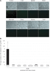
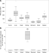
In ovo biodistribution of HAdV-5 particles in tumor-bearing fertilized chicken eggs. 5E9 eGFP-expressing, replication-incompetent HAdV-5 vector particles dissolved in 50 μL PBS were intravenously injected in human UM-SCC-11B tumor-bearing chicken eggs at E12. At E14, chicks were sacrificed and tumors and chick organs harvested. (A) Tumors and organs were homogenized, protein concentrations determined by measurement of the optical density at 280 nm, and the eGFP fluorescence intensity analyzed using a fluorospectrometer (n = 19). (B) Tumors and tissues were harvested and total DNA extracted. The vector genome copy number of 20 ng total DNA was quantified by qPCR and normalized with human β-actin copy numbers (n = 7). **p < 0.005. RFU, response fluorescence units.
Figure 6.
Figure 6.
Intratumoral virus replication but insufficient oncolysis. 5E9 eGFP-expressing, replication-competent HAdV-5wt virus or replication-incompetent HAdV-5 vector particles dissolved in 50 μL PBS were intravenously injected in human UM-SCC-11B tumor-bearing chicken eggs at E11. At E16, chicks were sacrificed and tumors harvested. (A) Six-micron cryosections of tumors were prepared and analyzed for eGFP-expressing cells by fluorescence microscopy. Fivefold magnification, representative data of n = 3. (B) Total DNA of tumors was extracted. The viral genome copy number of 20 ng total DNA was quantified by qPCR and normalized to human β-actin copy numbers (n = 14–15). (C) Tumors were weighed after excision (n = 17–18). Scale bar: 200 μm, ***p < 0.0005.

Similar articles

Cited by

References

    1. Bluming AZ, Ziegler JL. Regression of Burkitt's lymphoma in association with measles infection. Regres Burkitts Lymphoma Assoc Measles Infect 1971;105–106 - PubMed
    1. Russell L, Peng K-W. The emerging role of oncolytic virus therapy against cancer. Chin Clin Oncol 2018;7:16. - PMC - PubMed
    1. Murphy JB. Transplantability of tissues to the embryo of foreign species: its bearing on questions of tissue specificity and tumor immunity. J Exp Med 1913;17:482–493 - PMC - PubMed
    1. Murphy JB, Rous P. The behavior of chicken sarcoma implanted in the developing embryo. J Exp Med 1912;15:119–132 - PMC - PubMed
    1. Mullen JT, Donahue JM, Chandrasekhar S, et al. . Oncolysis by viral replication and inhibition of angiogenesis by a replication-conditional herpes simplex virus that expresses mouse endostatin. Cancer 2004;101:869–877 - PubMed