Review
. 2020 Sep 15:6:313-334.
doi: 10.1146/annurev-vision-121219-081716. Epub 2020 Jun 17.

Role of Feedback Connections in Central Visual Processing

Affiliations
Review

Role of Feedback Connections in Central Visual Processing

Farran Briggs. Annu Rev Vis Sci. .

Abstract

The physiological response properties of neurons in the visual system are inherited mainly from feedforward inputs. Interestingly, feedback inputs often outnumber feedforward inputs. Although they are numerous, feedback connections are weaker, slower, and considered to be modulatory, in contrast to fast, high-efficacy feedforward connections. Accordingly, the functional role of feedback in visual processing has remained a fundamental mystery in vision science. At the core of this mystery are questions about whether feedback circuits regulate spatial receptive field properties versus temporal responses among target neurons, or whether feedback serves a more global role in arousal or attention. These proposed functions are not mutually exclusive, and there is compelling evidence to support multiple functional roles for feedback. In this review, the role of feedback in vision will be explored mainly from the perspective of corticothalamic feedback. Further generalized principles of feedback applicable to corticocortical connections will also be considered.

Keywords: corticocortical; corticogeniculate; corticothalamic; feedback; spatial receptive field properties; temporal receptive field properties.


Figures
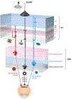
Figure 1
Figure 1
Feedforward and feedback parallel pathways in the early visual system of primates. Retinal ganglion cells conveying visual information to the dorsal lateral geniculate nucleus (LGN) of the thalamus are separated into three major parallel processing streams: the parvocellular (red), magnocellular (black), and koniocellular (blue) streams. These retinal ganglion cells (filled colored circles at bottom) project axons to separated parvocellular (pink), magnocellular (gray), and intercalated koniocellular (blue) layers of the LGN. LGN relay neurons (filled colored circles in LGN) display distinctive receptive field properties driven by their retinal inputs: red–green center–surround organization among parvocellular LGN neurons, On–Off (black–white) center–surround organization among magnocellular LGN neurons, and blue–yellow opponent organization among koniocellular neurons. The geniculocortical axons of LGN relay neurons target segregated layers within the primary visual cortex (V1): Parvocellular geniculocortical axons (red thin line) mainly target layer 4Cβ (pink); magnocellular geniculocortical axons (black thin line) mainly target layer 4Cα (gray); and koniocellular geniculocortical axons (blue thin line) target layer 1, the blobs in layer 2/3, and layer 4A. Geniculocortical recipient neurons in these V1 layers also display distinctive receptive field properties driven by their geniculocortical inputs: Parvocellular-recipient V1 neurons are simple cells with separable red–green receptive field subregions, magnocellular-recipient V1 neurons are simple and complex cells with orientation tuning and direction selectivity, and koniocellular-recipient V1 neurons are responsive to blue color. Corticogeniculate neurons located in layer 6 of V1 provide feedback from V1 to the LGN. Corticogeniculate neurons are also separated into the same parallel streams, and their receptive field properties reflect their feedforward stream-specific inputs. There are parvocellular-projecting corticogeniculate neurons (red pyramidal cell in upper layer 6 of V1), magnocellular-projecting corticogeniculate neurons (black pyramidal cell in lower layer 6), and koniocellular-projecting corticogeniculate neurons (blue tilted cell at the bottom of layer 6). The distributions of corticogeniculate axon terminals in the LGN (red, black, and blue downward cones) are broader compared to the distributions of retinogeniculate axon terminals in the LGN (red, black, blue upward cones). Similarly, the distribution of terminals from corticocortical feedback axons (top black cone) are also more broadly distributed relative to feedforward and local axon terminal distributions within V1. Corticocortical feedback originates in extrastriate visual areas such as the secondary visual cortex (V2) and the middle temporal area (MT), in which neurons are responsive to more complex visual features including texture and stimulus motion.

Similar articles

Cited by

References

LITERATURE CITED

    1. Alexander GM, Godwin DW. 2005. Presynaptic inhibition of corticothalamic feedback by metabotropic glutamate receptors. J. Neurophysiol 94:163–75 - PubMed
    1. Alitto HJ, Usrey WM. 2008. Origin and dynamics of extraclassical suppression in the lateral geniculate nucleus of the macaque monkey. Neuron 57:135–46 - PMC - PubMed
    1. Alonso J-M, Usrey WM, Reid RC. 1996. Precisely correlated firing in cells of the lateral geniculate nucleus. Nature 383:815–19 - PubMed
    1. Anderson JC, Da Costa NM, Martin KAC. 2009. The W cell pathway to cat primary visual cortex. J. Comp. Neurol 516:20–35 - PubMed
    1. Anderson JC, Martin KAC. 2009. The synaptic connections between cortical areas V1 and V2 in macaque monkey. J. Neurosci 29:11283–93 - PMC - PubMed

RELATED RESOURCES

    1. Briggs F, Usrey WM. 2011. Corticogeniculate feedback and parallel processing in the primate visual system. J. Physiol 589:33–40 - PMC - PubMed
    1. Briggs F, Usrey WM. 2013. Functional properties of cortical feedback to the primate lateral geniculate nucleus. In The New Visual Neurosciences, ed. Werner JS, Chalupa LM, pp. 315–22. Cambridge, MA: MIT Press
    1. Usrey WM, Alitto HJ. 2015. Visual functions of the thalamus. Annu. Rev. Vis. Sci 1:351–71 - PMC - PubMed

Publication types

LinkOut - more resources