. 2020 Jun 18;11(1):243.
doi: 10.1186/s13287-020-01766-9.

Novel venom-based peptides (P13 and its derivative-M6) to maintain self-renewal of human embryonic stem cells by activating FGF and TGFβ signaling pathways

Affiliations

Novel venom-based peptides (P13 and its derivative-M6) to maintain self-renewal of human embryonic stem cells by activating FGF and TGFβ signaling pathways

Rui Ma et al. Stem Cell Res Ther. .

Abstract

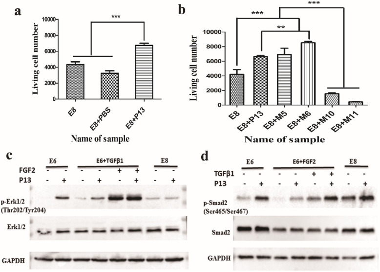
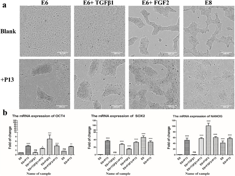
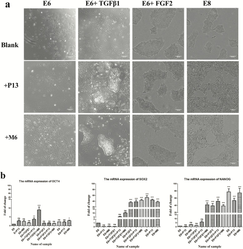
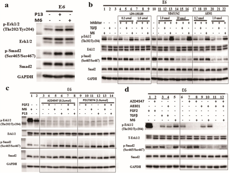
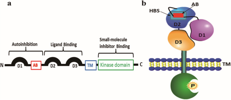
Background: In our previous study, a venom-based peptide named Gonearrestide (also named P13) was identified and demonstrated with an effective inhibition in the proliferation of colon cancer cells. In this study, we explored if P13 and its potent mutant M6 could promote the proliferation of human embryonic stem cells and even maintain their self-renewal.

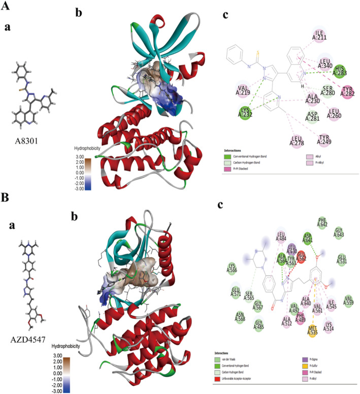
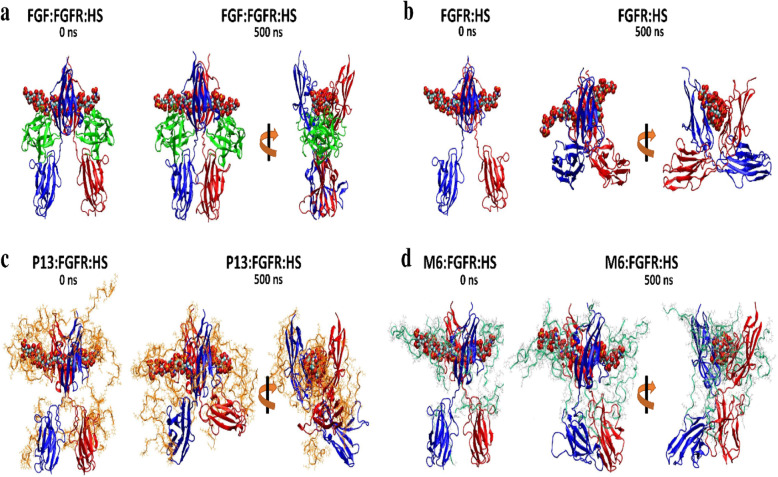
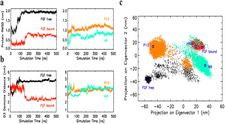
Methods: The structure-function relationship analysis on P13 and its potent mutant M6 were explored from the molecular mechanism of corresponding receptor activation by a series of inhibitor assay plus molecular and dynamics simulation studies.

Results: An interesting phenomenon is that P13 (and its potent mutant M6), an 18AA short peptide, can activate both FGF and TGFβ signaling pathways. We demonstrated that the underlying molecular mechanisms of P13 and M6 could cooperate with proteoglycans to complete the "dimerization" of FGFR and TGFβ receptors.

Conclusions: Taken together, this study is the first research finding on a venom-based peptide that works on the FGF and TGF-β signaling pathways to maintain the self-renewal of hESCs.

Keywords: Embryonic stem cell; Peptide modification; Pluripotency; Self-renewal; Venom peptide.


Conflict of interest statement

The authors declare no conflict of interest.

Figures

Fig. 1
Fig. 1
The survival ability and expression levels of p-Erk1/2 and p-Smad2 in human ES cells. a The survival ability of the human ES cells treated with venom peptide P13; the concentration of P13 was 200 μM/L; 1 × PBS was the solvent of the same volume used to dissolve the peptides. The statistical significances are marked by asterisks—*p < 0.05, **p < 0.01, and ***p < 0.001—between the two indicated groups. b The survival ability of the human ES cells treated with P13 and its mutants (M5, M6, M10, and M11); the concentration of P13 and its mutants was 200 μM/L. c The expression of the total/phosphorylation Erk1/2 in different culture conditions (E6, E6 + TGFβ1, and E8) when treated with P13 (left). d The expression of total/phosphorylation Smad2 in different culture conditions (E6, E6 + FGF2, and E8) when treated with P13 (right); the concentration of FGF2 was 100 μg/L, and the concentration of TGFβ1 was 2 μg/L.
Fig. 2
Fig. 2
The human ES cells’ morphology and self-renewal capacity when treated with P13. a These images of the human ES cells were taken after passaging five times (magnification, 10×); the concentration of P13 was 200 μM/L, and the blank represents the 1 × PBS solvent for dissolving the peptides. b The qPCR results of POU5 F1, SOX2, and NANOG of the human ES cells in different culture conditions and treated with P13
Fig. 3
Fig. 3
The human ES cells’ morphology and self-renewal ability when treated with M6. a The microscope images were taken after the human ES Cells were passaged five times, and P13 was used as a control (magnification, 10×); the concentration of P13 and M6 was 100 μM/L, and the blank represents the 1 × PBS solvent used to dissolve peptides. b The qPCR results of OCT4, SOX2, and NANOG of the human ES cells in different culture conditions treated with P13/M6
Fig. 4
Fig. 4
The location of P13 and its mutant M6, labeled by FITC, in the membrane of human ES cells. Confocal microscope figures with 100× oil magnification (a P13-FITC and b M6-FITC)
Fig. 5
Fig. 5
The expressions of p-Erk1/2 and p-Smad2 in the human ES cells treated with M6 without/with small molecular inhibitors. a The expression of total/phosphorylation Erk1/2 and total/phosphorylation Smad2 when treated with P13 and its mutant M6 without any inhibitor. b The expressions of the TGFβ signaling pathways after being treated with TGFβ receptor inhibitors (LDN-193189, SB431542, and A8301); the concentration of SB431542 was 1.0 and 10 μmol, and the concentration of A8301/LDN-193189 was 0.2 and 1.0 μmol. c The expression of the FGFR pathways after being treated with FGFR receptor inhibitors (AZD4547 and PD173074); the concentration of AZD4547 was 0.2 μmol, and the concentration of PD173074 was 1.0 μmol. d The expressions of the FGF and TGFβ signaling pathways after being treated with the FGFR and TGFβ receptor dual inhibitors (0.2 μmol AZD4547 and 1.0 μmol A8301)
Fig. 6
Fig. 6
The molecular modeling of the kinase domain treated with small molecular inhibitors. A The solid ribbon modeling of the FGFR kinase domain bound to AZD4547 based on the PDB ID 4WUN: (a) represents the 2D structure of AZD4547; (b) represents the AZD4547 bound to the pocket of the FGFR intracellular kinase domain; and (c) is a 2D model of the interaction between AZD4547 and FGFR in the active molecular window. B The solid ribbon modeling of the TGFβ receptor kinase domain bound to A8301 based on the PDB ID 3TZM: (a) represents the 2D structure of A8301; (b) shows the A8301 bound to the pocket of the TGFβ receptor intracellular kinase domain; and (c) is a 2D model of the interaction between A8301 and TGFβ in the active molecular window
Fig. 7
Fig. 7
Simulation snapshots of four types of the simulation system. a The crystal FGF:FGFR:HS complex. b The apo FGFR:HS complex. c The FGFR:HS complex with 20 chains of P13. d The FGFR:HS complex with 20 chains of M6. Few peptides that are not in contact with protein were removed for clarity
Fig. 8
Fig. 8
Analysis of structural changes of FGFR under different ligand conditions: FGF-free, FGF-bound, P13, and M6. a The root-mean-squared deviation (RMSD) of the protein backbone in reference to its initial structure. b The center-of-mass separation distance between the D3 domains of the two FGFR chains. c Two-dimensional projection of the four combined trajectories (FGF-free, FGF-bound, P13, and M6) on the first and second PCA modes computed from the coordinates of FGFR backbone atoms. The square symbols indicate positions of protein conformations in terms of PCA coordinates for the initial (all trajectories have the same initial protein conformation) and the final state (500 ns) of each trajectory. Color codes for trajectory: FGF-bound (red), FGF-free (black), P13 (orange), M6 (cyan)
Fig. 9
Fig. 9
Schematic diagram and autoinhibition model of FGFR. a Schematic diagram of FGFR: The “AB” in red stands for acid box, and TM represents the transmembrane domain. Kinase domain phosphorylates downstream molecules and can be inhibited by small-molecule inhibitors. The extracellular region of FGFR is involved in receptor autoinhibition, and the ligand-binding is indicated. The N- and C-terminals are labeled N and C, respectively. b Autoinhibition model of FGFR: The HBS is the heparin-binding site. AB binds to the HBS of D2 to inhibit the activation of FGFR. Figure source adopted from Mohammadi et al. [47, 48] and modified by Rui MA

Similar articles

Cited by

References

    1. King GF. Venoms as a platform for human drugs: translating toxins into therapeutics. Expert Opin Biol Ther. 2011;11(11):1469–1484. - PubMed
    1. Ma R, Mahadevappa R, Kwok HF. Venom-based peptide therapy: insights into anti-cancer mechanism. Oncotarget. 2017;8(59):100908–100930. - PMC - PubMed
    1. Li B, Lyu P, Xi XP, Ge LL, Mahadevappa R, Shaw C, Kwok HF. Triggering of cancer cell cycle arrest by a novel scorpion venom-derived peptide-Gonearrestide. J Cell Mol Med. 2018;22(9):4460–4473. - PMC - PubMed
    1. King GF, Escoubas P, Nicholson GM. Peptide toxins that selectively target insect Na-V and Ca-V channels. Channels. 2008;2(2):100–116. - PubMed
    1. de la Vega RC R, Possani LD. Overview of scorpion toxins specific for Na+ channels and related peptides: biodiversity, structure-function relationships and evolution. Toxicon. 2005;46(8):831–844. - PubMed

Publication types

LinkOut - more resources