. 2020 Jul 16;27(7):839-849.e4.
doi: 10.1016/j.chembiol.2020.05.013. Epub 2020 Jun 16.

tRNAArg-Derived Fragments Can Serve as Arginine Donors for Protein Arginylation

Affiliations

tRNAArg-Derived Fragments Can Serve as Arginine Donors for Protein Arginylation

Irem Avcilar-Kucukgoze et al. Cell Chem Biol. .

Abstract

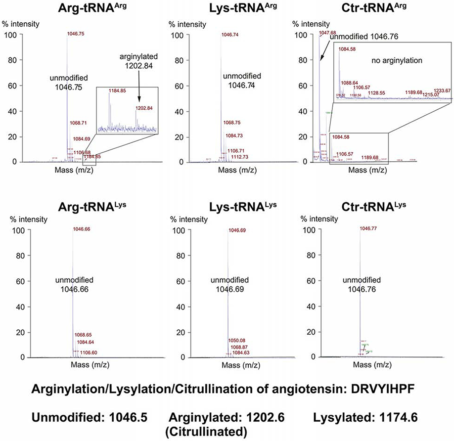
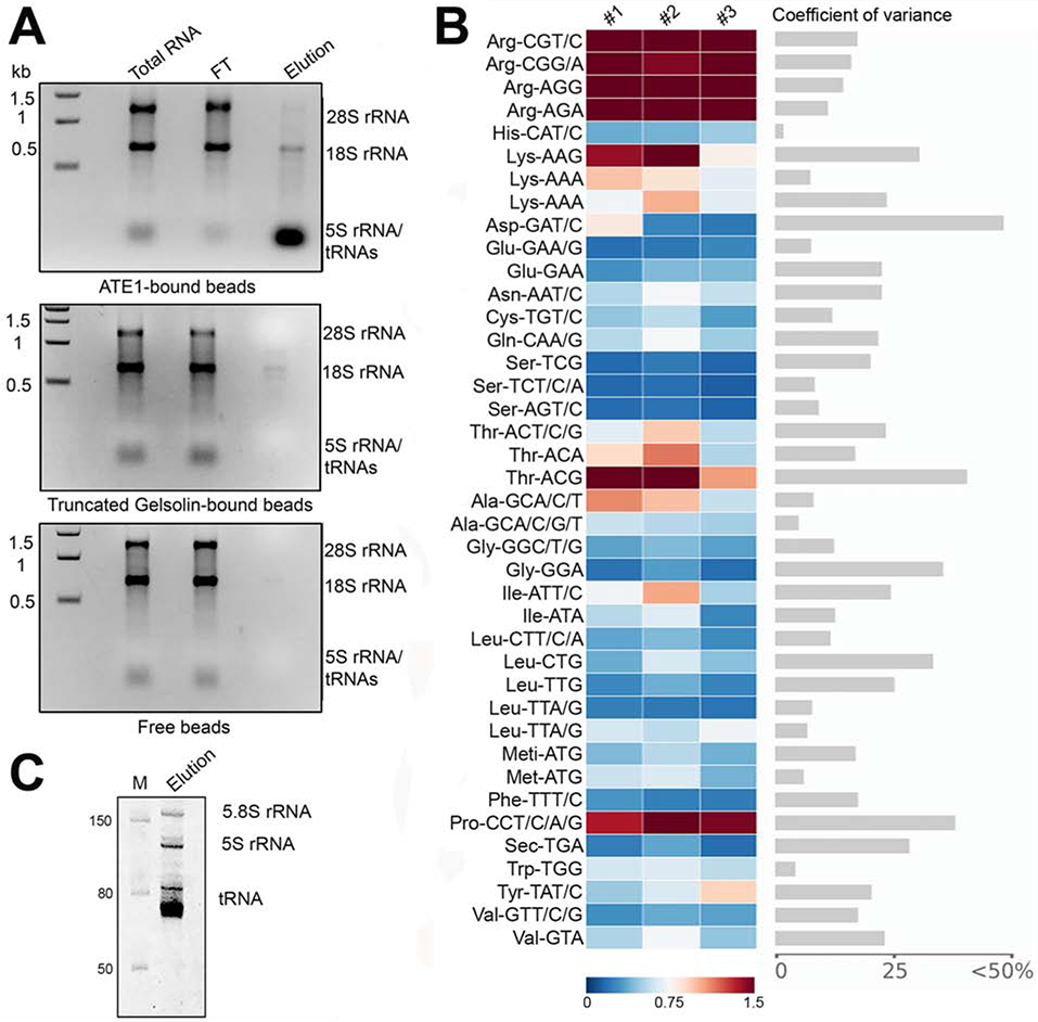
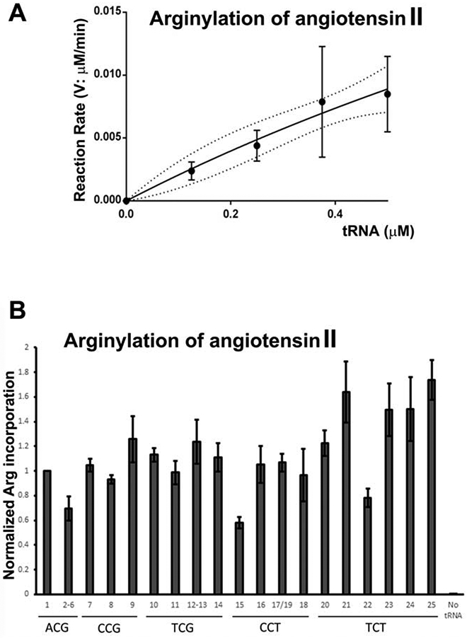
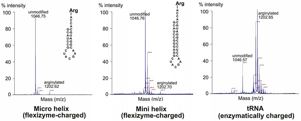
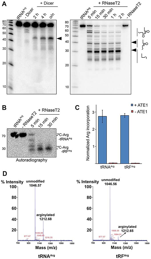
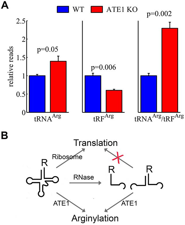
Arginyltransferase ATE1 mediates posttranslational arginylation and plays key roles in multiple physiological processes. ATE1 utilizes arginyl (Arg)-tRNAArg as the donor of Arg, putting this reaction into a direct competition with the protein synthesis machinery. Here, we address the question of ATE1- Arg-tRNAArg specificity as a potential mechanism enabling this competition in vivo. Using in vitro arginylation assays and Ate1 knockout models, we find that, in addition to full-length tRNA, ATE1 is also able to utilize short tRNAArg fragments that bear structural resemblance to tRNA-derived fragments (tRF), a recently discovered class of small regulatory non-coding RNAs with global emerging biological role. Ate1 knockout cells show a decrease in tRFArg generation and a significant increase in the ratio of tRNAArg:tRFArg compared with wild type, suggesting a functional link between tRFArg and arginylation. We propose that generation of physiologically important tRFs can serve as a switch between translation and protein arginylation.

Keywords: arginylation; tRF; tRNA.


Conflict of interest statement

Declaration of Interests The authors declare no competing interests.

Figures

Figure 1.
Figure 1.. ATE1-mediated arginylation is highly specific toward Arg-tRNAArg.
MALDI-TOF chromatograms of angiotensin II. Masses of unmodified and arginylated angiotensin are marked. Only tRNAArg charged with Arg is able to mediate arginylation.
Figure 2.
Figure 2.. ATE1 selectively binds to tRNA.
A, Gels of total RNA used as pulldown input, flow through (FT) and RNA eluate from His-tagged ATE1 immobilized on Ni-NTA beads (top), His-tagged gelsolin fragment immobilized on Ni-NTA beads (middle) and Ni-NTA beads alone (bottom). B, Comparative microarrays of tRNAs bound to ATE1 vs total tRNA of mouse liver summary of three biological replicates. Arrays are highly reproducible (Fig. S2). tRNA probes are depicted with their codon and the corresponding amino acid; Meti-ATG, initiator tRNAMet. Two different probes recognizing different tRNALeu (Leu-UAA) and tRNALys (Lys-UUU) isodecoders that pair to the same codon TTA/G-Leu or AAA-Lys codon but differ in their sequence outside the anticodon were used on the arrays. Data are shown as fold enrichment (gradient ruler at the bottom). Significantly enriched tRNAs in the ATE1 pull down compared to the total liver tRNAs were selected based on the confidence intervals of covariance analysis (an example between replicate 1 and 2 is included). C, Eluted RNA sample in denaturing 10% polyacrylamide gel. M, RNA marker.
Figure 3.
Figure 3.. ATE1 is able to utilize all mouse tRNAArg species.
A, Rate of the arginylation reaction as a function of E. coli Arg-tRNAArg in the presence of excess of angiotensin II substrate B, Normalized Arg incorporation into angiotensin II using various pre-charged mouse Arg-tRNAArg isoacceptors and isodecoders. No tRNA, control without adding tRNA (see Table S1 for tRNA gene names and sequences, corresponding to the numbers on the chart).
Figure 4.
Figure 4.. tRNAArg acceptor stem-like structures charged with Arg can mediate arginylation.
MALDI-TOF chromatograms of angiotensin II, arginylated in vitro using flexizyme-charged Arg-mini- and microhelix (left and middle panel), or RARS-charged Arg-tRNAArg. Masses of unmodified and arginylated angiotensin are marked. Structures of micro- and minihelix tRNAArg used in the reaction are shown on each chromatogram. The higher arginylated peak intensity in the tRNA-mediated reaction is likely due to the fact that this is a fully enzymatic reaction, where RARS present in the mixture continually charges tRNA for the arginylation reaction. Notably, the peak height/intensity in this method does not directly reflect the abundance of the peptide (Szajli et al., 2008).
Figure 5.
Figure 5.. tRNAArg-derived fragments mediate arginylation.
A, tRNA cleavage pattens by Dicer and RNase T2. The arrows show the cleavage products. B, Autoradiogram of 14C-Arg-tRNAArg cleaved with RNase T2. C, normalized incorporation of 3H-Arg into angiotensin II performed with 3H-Arg-tRNAArg or 3H-Arg-tRFArg. Data are mean ± SEM of three biological replicates. D. MALDI-TOF chromatogram of angiotensin II arginylated using purified pre-charged 13C15N-Arg-tRNAArg or 13C15N-Arg-tRFArg. Notably, the peak height/intensity in this method does not directly reflect the abundance of the peptide (Szajli et al., 2008).
Figure 6.
Figure 6.. ATE1 deletion alters the ratio of tRNAArg to tRFArg in cells.
A. Ratios of tRNAArg/tRFArg in the total RNA preparations from wild type (WT) and ATE1 knockout (ATE1 KO) mouse embryonic fibroblasts, analyzed in 3 biological replicates. Error bars represent SEM, Welch’s one-tailed T-test was used for calculating statistical significance. B, Hypothetical model of the arginylation and translation dependent tRNAArg/tRFArg balance.

Similar articles

Cited by

References

    1. Andersen KL, and Collins K (2012). Several RNase T2 enzymes function in induced tRNA and rRNA turnover in the ciliate Tetrahymena. Molecular biology of the cell 23, 36–44. - PMC - PubMed
    1. Anderson P, and Ivanov P (2014). tRNA fragments in human health and disease. FEBS letters 588, 4297–4304. - PMC - PubMed
    1. Barciszewska MZ, Erdmann VA, and Barciszewski J (1996). Ribosomal 5S RNA: tertiary structure and interactions with proteins. Biological reviews of the Cambridge Philosophical Society 71, 1–25. - PubMed
    1. Batsios P, Ishikawa-Ankerhold HC, Roth H, Schleicher M, Wong CCL, and Muller-Taubenberger A (2019). Ate1-mediated posttranslational arginylation affects substrate adhesion and cell migration in Dictyostelium discoideum. Molecular biology of the cell 30, 453–466. - PMC - PubMed
    1. Beckert B, Turk M, Czech A, Berninghausen O, Beckmann R, Ignatova Z, Plitzko JM, and Wilson DN (2018). Structure of a hibernating 100S ribosome reveals an inactive conformation of the ribosomal protein S1. Nature microbiology 3, 1115–1121. - PubMed

Publication types