. 2020 Aug 24;54(4):455-470.e5.
doi: 10.1016/j.devcel.2020.05.027. Epub 2020 Jun 17.

DDX3X Suppresses the Susceptibility of Hindbrain Lineages to Medulloblastoma

Affiliations

DDX3X Suppresses the Susceptibility of Hindbrain Lineages to Medulloblastoma

Deanna M Patmore et al. Dev Cell. .

Abstract

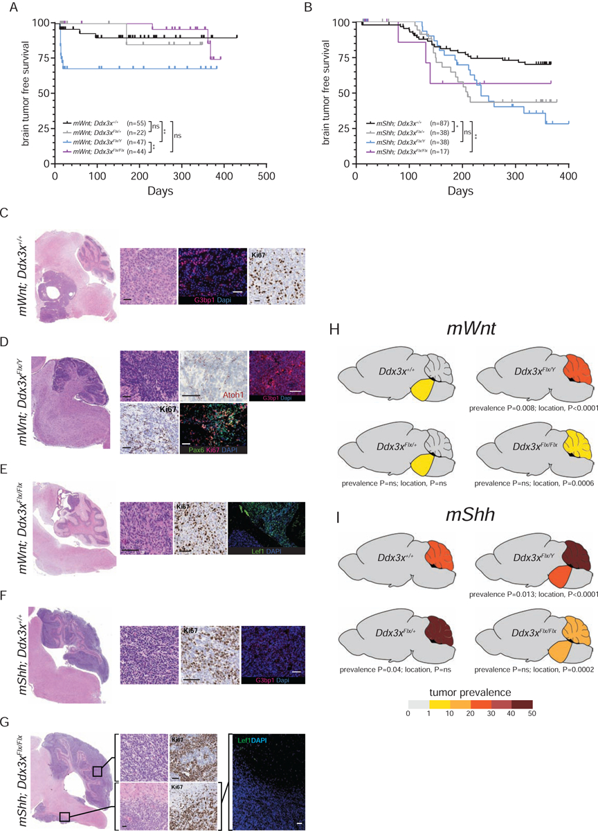
DEAD-Box Helicase 3 X-Linked (DDX3X) is frequently mutated in the Wingless (WNT) and Sonic hedghog (SHH) subtypes of medulloblastoma-the commonest malignant childhood brain tumor, but whether DDX3X functions as a medulloblastoma oncogene or tumor suppressor gene is not known. Here, we show that Ddx3x regulates hindbrain patterning and development by controlling Hox gene expression and cell stress signaling. In mice predisposed to Wnt- or Shh medulloblastoma, Ddx3x sensed oncogenic stress and suppressed tumor formation. WNT and SHH medulloblastomas normally arise only in the lower and upper rhombic lips, respectively. Deletion of Ddx3x removed this lineage restriction, enabling both medulloblastoma subtypes to arise in either germinal zone. Thus, DDX3X is a medulloblastoma tumor suppressor that regulates hindbrain development and restricts the competence of cell lineages to form medulloblastoma subtypes.

Keywords: DDX3X; HOX genes; inflammasome; medulloblastoma; stress granule; tumor suppressor gene.


Conflict of interest statement

Declaration of Interests The authors declare no competing interests.

Figures

Figure 1.
Figure 1.. Ddx3x regulates hindbrain development.
A. Western blot of Ddx3x and actin loading control expression in Cre-recombined mouse NPCs of the indicated genotype. B. Phenotype-free survival of Blbp-Cre+/−;Ddx3xFlx/Flx (n=55), Blbp-Cre+/−;Ddx3xFlx/+ (n=29) and Blbp-Cre+/−;Ddx3xFlx/Y mice (n=31). C. Heatmaps reporting the log-fold difference in the indicated histological parameters quantified by automated photo-micromorphometry. Analyses were performed on ≥10 mice per time point for each indicated variable and genotype. D. Photomicrographs of morphology (hematoxylin and eosin stain) and Ddx3x RNA expression (in situ hybridization) in developing hindbrains of the indicated genotype (unless indicated scale bars=100μm). E. Microanatomy of the cerebellar cortex cells and associated markers used in F-H are: EGL=external germinal layer; IGL=internal granule cell layer; BG=Bergman glia; PC=Purkinje cell; SC=stellate cell. F. Photomicrographs of morphology (hematoxylin and eosin stain) of the P13 cerebellar cortex in the indicated mouse genotype (scale bars=100μm). G, H. In situ hybridization (Atoh1) and immunofluorescence of the indicated cell markers in P13 mouse cerebellar cortex of the indicated genotype. I. Ddx3y fragments per kilobase of transcript per million mapped reads in mouse hindbrain (n=3 to 5 per genotype, sex and age; *=p<0.05, Mann-Whitney). See also Figure S1.
Figure 2.
Figure 2.. Ddx3x regulates tissue patterning and stress response in the hindbrain.
A. Principal components analysis of RNAseq profiles generated from hindbrains isolated from the indicated mice (≥5 hindbrains per timepoint and genotype). B. Centre, heat map of the log-fold difference of expression of the indicated genes in hindbrains of mice with the indicated genotypes relative to Blbp-Cre+/−;Ddx3x+/+ controls (≥5 hindbrains per timepoint and genotype at the indicated developmental time points). Left, Cytoscape™ plots identifying gene sets with related functions that are significantly up- or down-regulated in Blbp-Cre+/−;Ddx3xFlx/Flx hindbrains relative to Blbp-Cre+/−;Ddx3x+/+ controls at the indicated developmental stage. Right, in situ hybridization of the indicated genes in Blbp-Cre+/−;Ddx3xFlx/Flx and Blbp-Cre+/−;Ddx3x+/+ hindbrains. Cartoons indicate the location of the higher power images (scale bars=100μm). See also Figure S2.
Figure 3.
Figure 3.. Ddx3x is a medulloblastoma tumor suppressor gene.
Tumor-free survival curves of mice predisposed to develop either Wnt- (A) or Shh-medulloblastoma (B) harboring the indicated Ddx3x alleles. Numbers of mice ‘n’ are shown for each cohort. ns=not significant, *=p<0.05, **=p<0.005, Log-Rank statistic. Photomicrographs of low and high-power images of tumors arising in mice predisposed to develop Wnt- (C-E) or Shh-medulloblastomas (F,G) harboring the indicated Ddx3x alleles. High power images reveal tumor morphology (hematoloxylin and eosin), proliferation (Ki67 immunohistochemical stain), or immunofluorescence of the indicated markers. T=tumor, (scale bars=100μm). Cartoons depicting the incidence and distribution (brainstem or cerebellum) of tumors arising in mice predisposed to develop either Wnt- (H) or Shh-medulloblastoma (I) harboring the indicated Ddx3x alleles. Mouse numbers same as in (A and B). See also Figure S3.
Figure 4.
Figure 4.. Ddx3x restricts cell lineage origins of Wnt-medulloblastoma.
A. Principal component analysis of RNAseq profiles generated from 30 mouse tumors (shown in B) arising in mice predisposed to develop Wnt- or Shh-medulloblastomas harboring the indicated Ddx3x alleles. B. Unsupervised hierarchical clustering of RNAseq profiles of 30 tumors shown in (A). Heatmap shows the log2 normalized expression values of the indicated genes. Gene set classification is shown left, including signature genes of WNT and SHH medulloblastoma. C. Cytoscape™ plot identifying gene sets with related functions that are significantly up- or down-regulated in mWnt;Ddx3xFlx/Y relative to mWnt;Ddx3x+/+ tumors shown in A and B. D. Tumor growth recorded as bioluminescence in mice implanted with cells isolated from cerebella of the indicated genotype (number of mice implanted with each cell type is shown ‘n’; ***=p<0.0005, Mann-Whitney). E. Photomicrographs of tumor (T) arising in the cerebellum of a recipient mice implanted with cerebellar cells isolated from mWnt;Ddx3xFlx/Y mice. Images reveal tumor morphology (hematoloxylin and eosin), proliferation (Ki67 immunohistochemical stain), or Lef1 immunofluorescence (scale bars=100μm). See also Figure S4.
Figure 5.
Figure 5.. Ddx3x suppresses oncogenic Wnt-signaling in the developing cerebellum.
Heatmaps reporting the log-fold difference (LFD) in expression of the indicated genes (A) or False Discovery Rate of the indicated pathway up- or down-regulation (B) in premalignant cerebellar isolated from mice predisposed to develop either Wnt- (left) or Shh-medulloblastoma (right) harboring the indicated Ddx3x alleles at the indicated developmental time points relative to Blbp-Cre+/−;Ddx3x+/+ controls (≥5 cerebellar/genotype/time point). Fold change in Ki67 (C), p21 (D) and cleaved Caspase 3 (E) immunohistochemistry in the mice shown in (A and B) relative to Blbp-Cre+/−;Ddx3x+/+ controls (n≥10). The number ‘n’ of brains analyzed in each assay is shown above each bar. *=p<0.05; **=p<0.005; ***=p<0.0005, Mann-Whitney. F. Exemplary immunohistochemical stains in P5 cerebella of the indicated mice; scale bar = 50μm.
Figure 6.
Figure 6.. Single cell RNAseq of human DDX3Xmut and DDX3Xwt WNT-medulloblastoma.
A. UMAP of single cell RNAseq profiles generated from human DDX3Xmut (n=2) and DDX3Xwt (n=3) WNT-medulloblastoma. B. Volcano plots of -log10 p-values versus log fold difference expression of genes in the human DDX3Xmut and DDX3Xwt WNT-medulloblastomas in (A). Coloured genes in each plot belong to the cell pathway with associated FDR enrichment (top left). Exemplar genes are indicated; those marked with an asterix were also upregulated in mouse Blpb-Cre+/−;mWnt;Ddx3xFlx/Y cerebella tumors. See also Figure 4C and S5.
Figure 7.
Figure 7.. Cartoon depicting the proposed role of Ddx3x in sensing oncogenic stress signals in the hindbrain.
In the context of wild-type Ddx3x shown left, oncogenic Wnt and Shh-signals in the lower and upper rhombic lip, respectively are relatively well tolerated. This provokes only a modest oncogenic stress response and fails to trigger a major inflammasome-pyroptotic death signal, markedly increasing the probability of forming medulloblastoma. Conversely, activation of oncogenic Wnt and Shh-signals in the upper and lower rhombic lip, respectively in which they are not primary mitogens, provokes a massive pyroptotic and tumor suppressing response. In the context of mutant Ddx3x shown right, this tumor suppressing pyroptotic cell death response is lost, allowing Wnt and Shh-medulloblastomas to form in either the upper or lower rhombic lips.

Comment in

References

    1. Anderson P, Kedersha N, and Ivanov P (2015). Stress granules, P-bodies and cancer. Biochim Biophys Acta 1849, 861–870. - PMC - PubMed
    1. Brai A, Martelli F, Riva V, Garbelli A, Fazi R, Zamperini C, Pollutri A, Falsitta L, Ronzini S, Maccari L, et al. (2019). DDX3X Helicase Inhibitors as a New Strategy To Fight the West Nile Virus Infection. J Med Chem 62, 2333–2347. - PubMed
    1. Dai H, Goto Y-I, and Itoh M (2017). Insulin-Like Growth Factor Binding Protein-3 Deficiency Leads to Behavior Impairment with Monoaminergic and Synaptic Dysfunction. Am J Pathol 187, 390–400. - PubMed
    1. Demir MK, Yapıcıer Ö, Mert B, Alshareefi W, and Bozbuğa M (2019). Primary Sonic Hedgehog-activated dorsal brainstem medulloblastoma and ipsilateral cerebellar atrophy in an adult. Neuroradiol J, 1971400919892824–1971400919892824. - PMC - PubMed
    1. Di Giannatale A, Carai A, Cacchione A, Marrazzo A, Dell’Anna VA, Colafati GS, Diomedi-Camassei F, Miele E, Po A, Ferretti E, et al. (2016). Anomalous vascularization in a Wnt medulloblastoma: a case report. BMC Neurol 16, 103–103. - PMC - PubMed

Publication types