Review
. 2020 Aug:197:108089.
doi: 10.1016/j.exer.2020.108089. Epub 2020 Jun 15.

Corneal wound healing

Affiliations
Review

Corneal wound healing

Steven E Wilson. Exp Eye Res. 2020 Aug.

Abstract

The corneal wound healing response is typically initiated by injuries to the epithelium and/or endothelium that may also involve the stroma. However, it can also be triggered by immune or infectious processes that enter the stroma via the limbal blood vessels. For mild injuries or infections, such as epithelial abrasions or mild controlled microbial infections, limited keratocyte apoptosis occurs and the epithelium or endothelium regenerates, the epithelial basement membrane (EBM) and/or Descemet's basement membrane (DBM) is repaired, and keratocyte- or fibrocyte-derived myofibroblast precursors either undergo apoptosis or revert to the parent cell types. For more severe injuries with extensive damage to EBM and/or DBM, delayed regeneration of the basement membranes leads to ongoing penetration of the pro-fibrotic cytokines transforming growth factor (TGF) β1, TGFβ2 and platelet-derived growth factor (PDGF) that drive the development of mature alpha-smooth muscle actin (SMA)+ myofibroblasts that secrete large amounts of disordered extracellular matrix (ECM) components to produce scarring stromal fibrosis. Fibrosis is dynamic with ongoing mitosis and development of SMA + myofibroblasts and continued autocrine-or paracrine interleukin (IL)-1-mediated apoptosis of myofibroblasts and their precursors. Eventual repair of the EBM and/or DBM can lead to at least partial resolution of scarring fibrosis.

Keywords: Cornea; Corneal fibroblasts; Fibrocytes; Fibrosis; Infection; Injury; Interleukin-1; Keratocyte apoptosis; Myofibroblasts; PDGF; Scarring. corneal; Stromal-epithelial interactions; TGF beta; Wound healing.


Conflict of interest statement

Proprietary interest statement

The author doesn’t have any commercial or proprietary interests in this study.

Figures

Fig. 1.
Fig. 1.
The original images illustrating keratocyte apoptosis in an adult Balb/C mouse cornea after epithelial scrape injury. A. Terminal deoxynucleotidyl transferase-dUTP nick end labeling (TUNEL) assay performed to detect anterior keratocytes with fragmented DNA characteristic of apoptosis (arrowheads) at four hours after epithelial debridement using a peroxidase-based TUNEL assay and darkfield light microscopy. 200X mag. B. Confirmatory transmission electron microscopy at one hour after epithelial debridement shows a keratocyte with chromatin condensation (C) and cytoplasmic contents of the cell in apoptotic bodies (arrowheads) that disperse into the surrounding stroma. 25,000X. Reprinted with permission from Wilson SE, et al. Exp. Eye Res. 1996;62:325–8.
Fig. 2.
Fig. 2.
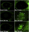
The influx of large numbers of bone marrow-derived cells into the corneas of chimeric mice with GFP-labeled bone marrow-derived cells after epithelial scrape injury. Few GFP+ cells were detected in control corneas that did not have epithelial scrape injuries (Con 0 is unwounded, Con 24 and Con 72 are unwounded at 24 and 72 hours, respectively). Similarly, few GFP+ cells are detected immediately following epithelial scrape (SCR 0 min). However, by one day after epithelial scrape injury (SCR 24 hrs), and continuing at 3 days after epithelial scrape injury (SCR 72 hrs), thousands of bone marrow-derived cells entered the cornea. Some GFP+ bone marrow-derived cells were detected in the corneas up to the final observation point of 10 days. Mag 10X. Reprinted with permission from Wilson et al. Invest. Ophthalmol. Vis. Sci, 2004;45:2201–2211.
Fig. 3.
Fig. 3.
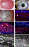
Fibrosis and myofibroblasts. A. At one month after −9 diopter PRK in the rabbit fibrosis (scarring or “late haze”) is present within the arrows, and is most dense on the left side of the cornea. B. At six months after penetrating keratoplasty in the human, fibrosis (arrows) is present within the donor-recipient interface and helps to hold the transplant in position once the sutures are removed. Immunohistochemistry performed on such corneas reveals myofibroblasts in the donor-recipient interface (not shown). C. At one month after severe Pseudomonas aeruginosa keratitis in the rabbit there is dense fibrosis (scarring) causing opacity of the entire cornea. Neovascularization (arrowheads) of the stroma can be noted. D. Immunohistochemistry for the myofibroblast marker α-smooth muscle actin (SMA) in a rabbit cornea at one month after −9D PRK shows a layer of myofibroblasts (arrows) in the anterior stroma. e is epithelium. Note there is artifactual dissociation of the epithelium from the myofibroblast-filled anterior stroma that occurred during sectioning of the cornea since the mature EBM had not regenerated. Mag. 200X. E. At one month after severe antibiotic-treated Pseudomonas aeruginosa keratitis in a rabbit, approximately 80% of the stroma is filled with SMA+ myofibroblasts (arrowheads). The infection did not spread completely through the stroma and there is normal stroma (*) with keratocytes just anterior to the endothelium. e is epithelium. Mag. 200X. F. In another rabbit cornea that had severe antibiotic-treated Pseudomonas aeruginosa keratitis at two months after infection, the anterior half of the stroma has very few SMA+ myofibroblasts, with the SMA present at neovascularization (arrows) being pericytes. In the posterior stroma, however, in this eye where the infection extended through the cornea and destroyed DBM and the endothelium, SMA+ myofibroblasts (arrowheads) fill the posterior stroma. e is epithelium. Mag 200X. G. Transmission electron microscopy (TEM) of the same cornea shown in E shows no epithelial basement membrane (EBM) lamina lucida or lamina densa beneath the epithelium (e). The anterior stroma is packed with stacked myofibroblasts (arrows) filled with rough endoplasmic reticulum. The stroma (s) between the myofibroblasts is disorganized. Mag. 25,000X. H. TEM of the same cornea shown in F shows EBM (arrows) with lamina lucida and lamina densa beneath the epithelium (e). Myofibroblasts have disappeared from the anterior stroma and the stromal matrix (s) is more organized, but not yet normal. Mag. 25,000X. C and E to H reprinted with permission from Marino GK, et al. Exp. Eye Res. 2017;161:101–105.
Fig. 4.
Fig. 4.
IL-1α expression in stromal cells at one month after −9D PRK in rabbits. A. DAPI staining to show nuclei in all cells at one month after PRK in a rabbit. B. IHC for the myofibroblasts marker SMA. Arrows indicate SMA+ myofibroblasts in the anterior stroma. C. IHC for IL-1α. Many anterior stromal cells produce IL-1α (arrows). D. Overlay of B and C shows that some of the stromal cells producing IL-1α are SMA+ myofibroblasts (arrows) but many surrounding cells that could be keratocytes, corneal fibroblasts or bone marrow-derived cells are also producing IL-1α. Mag 200X.

Similar articles

Cited by

References

    1. Ambrósio R Jr., Kara-José N, Wilson SE, 2009. Early keratocyte apoptosis after epithelial scrape injury in the human cornea. Exp. Eye Res 89, 597–9. - PMC - PubMed
    1. Barbosa FL, Chaurasia S, Cutler A, Asosingh K, Kaur H, de Medeiros F, Agrawal V, Wilson SE, 2010a. Corneal myofibroblast generation from bone marrow-derived cells. Exp. Eye Res 91, 92–6. - PMC - PubMed
    1. Barbosa FL, Chaurasia S, Kaur H, de Medeiros FW, Agrawal V, Wilson SE 2010b. Stromal interleukin-1 expression in the cornea after haze-associated injury. Exp. Eye Res 91, 456–61. - PMC - PubMed
    1. Barbosa FL, Lin M, Santhiago MR, Singh V, Agrawal V, Wilson SE, 2012. Interleukin-1 receptor role in the viability of corneal myofibroblasts. Exp. Eye Res 96, 65–9. - PMC - PubMed
    1. Behrens DT, Villone D, Koch M, Brunner G, Sorokin L, Robenek H, Bruckner-Tuderman L, Bruckner P, Hansen U, 2012. The epidermal basement membrane is a composite of separate laminin- or collagen IV-containing networks connected by aggregated perlecan, but not by nidogens. J. Biol. Chem 287, 18700–18709. - PMC - PubMed

Publication types