Review
. 2020 Sep:73:109696.
doi: 10.1016/j.cellsig.2020.109696. Epub 2020 Jun 15.

Characteristics of circular RNAs generated by human Survival Motor Neuron genes

Affiliations
Review

Characteristics of circular RNAs generated by human Survival Motor Neuron genes

Eric W Ottesen et al. Cell Signal. 2020 Sep.

Abstract

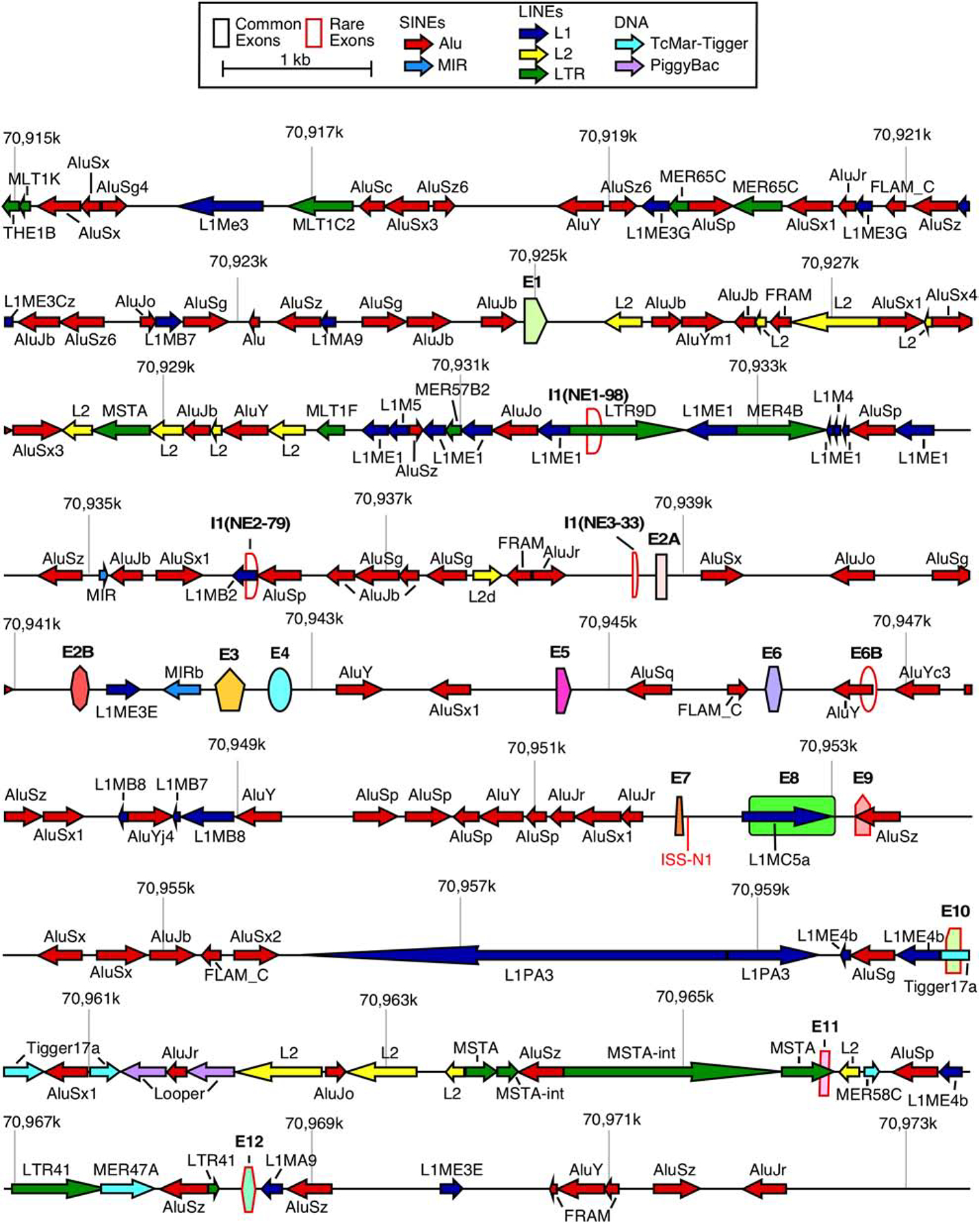
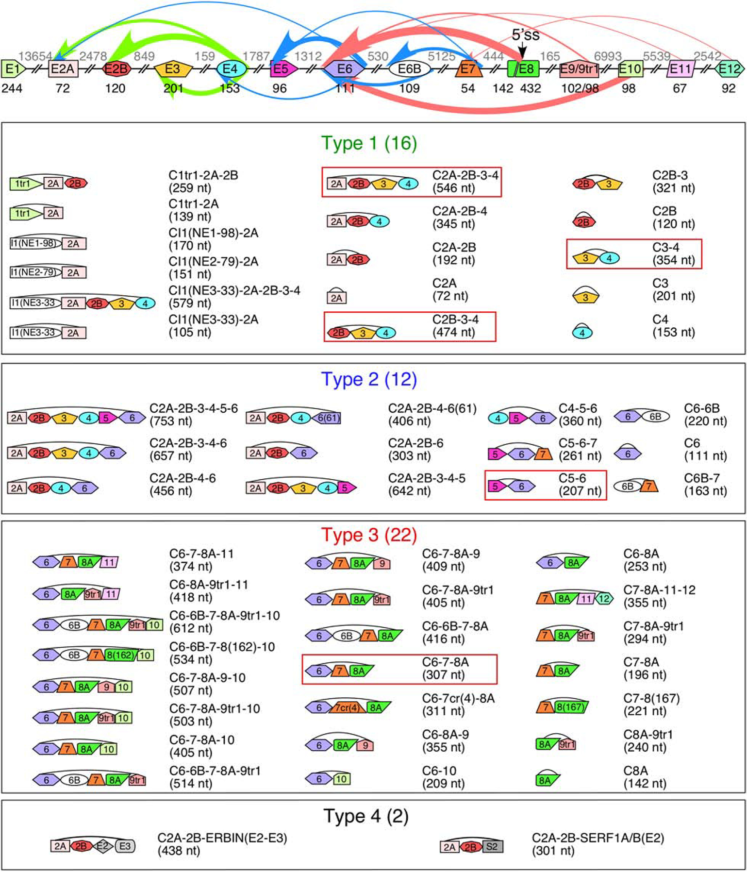
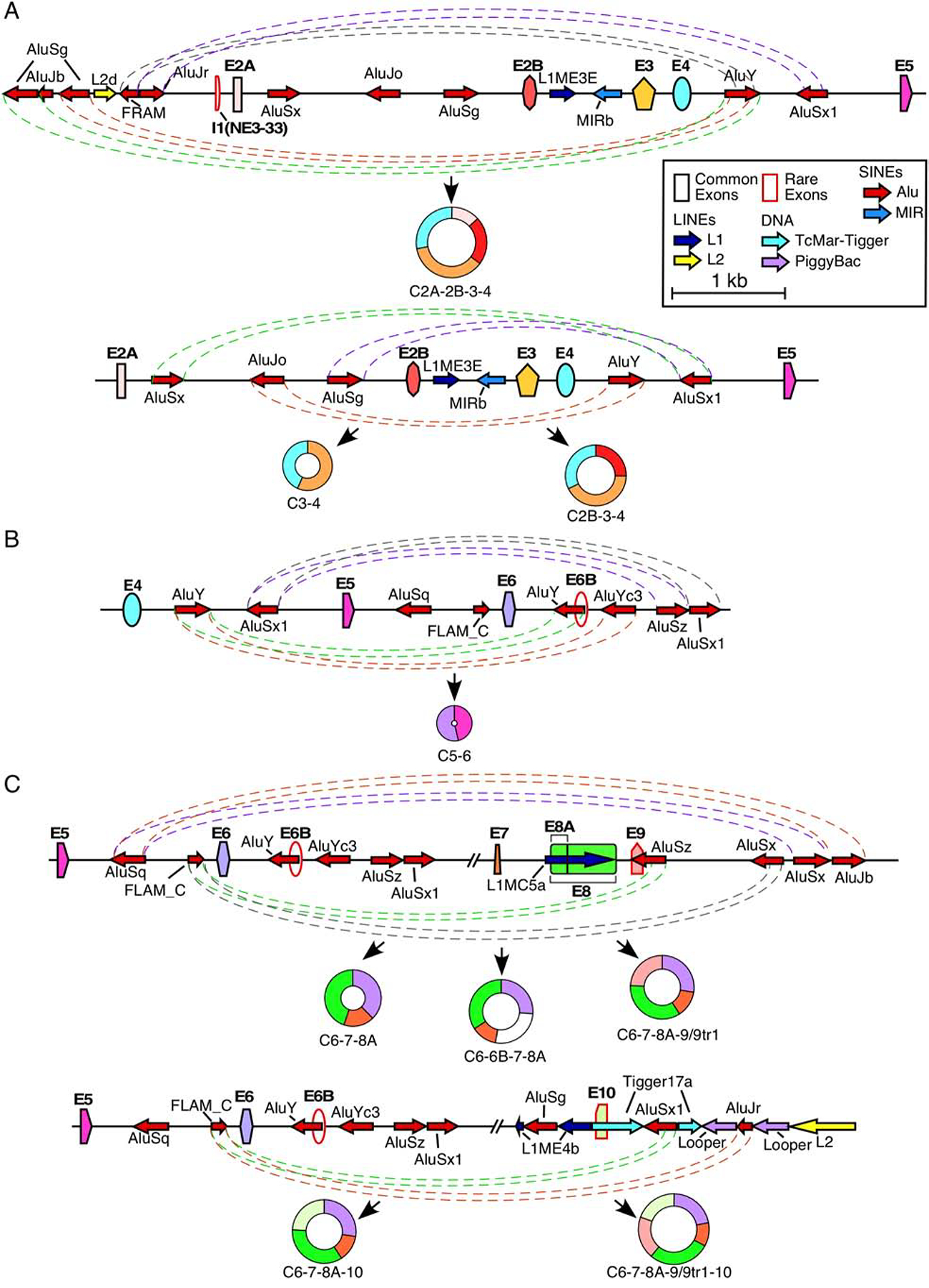
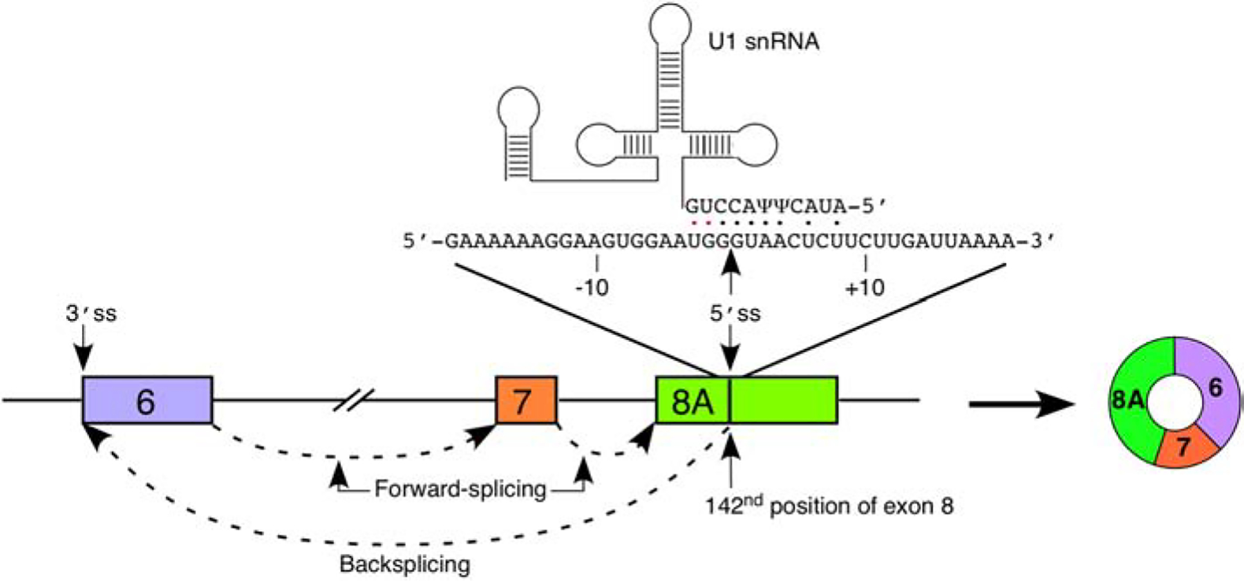
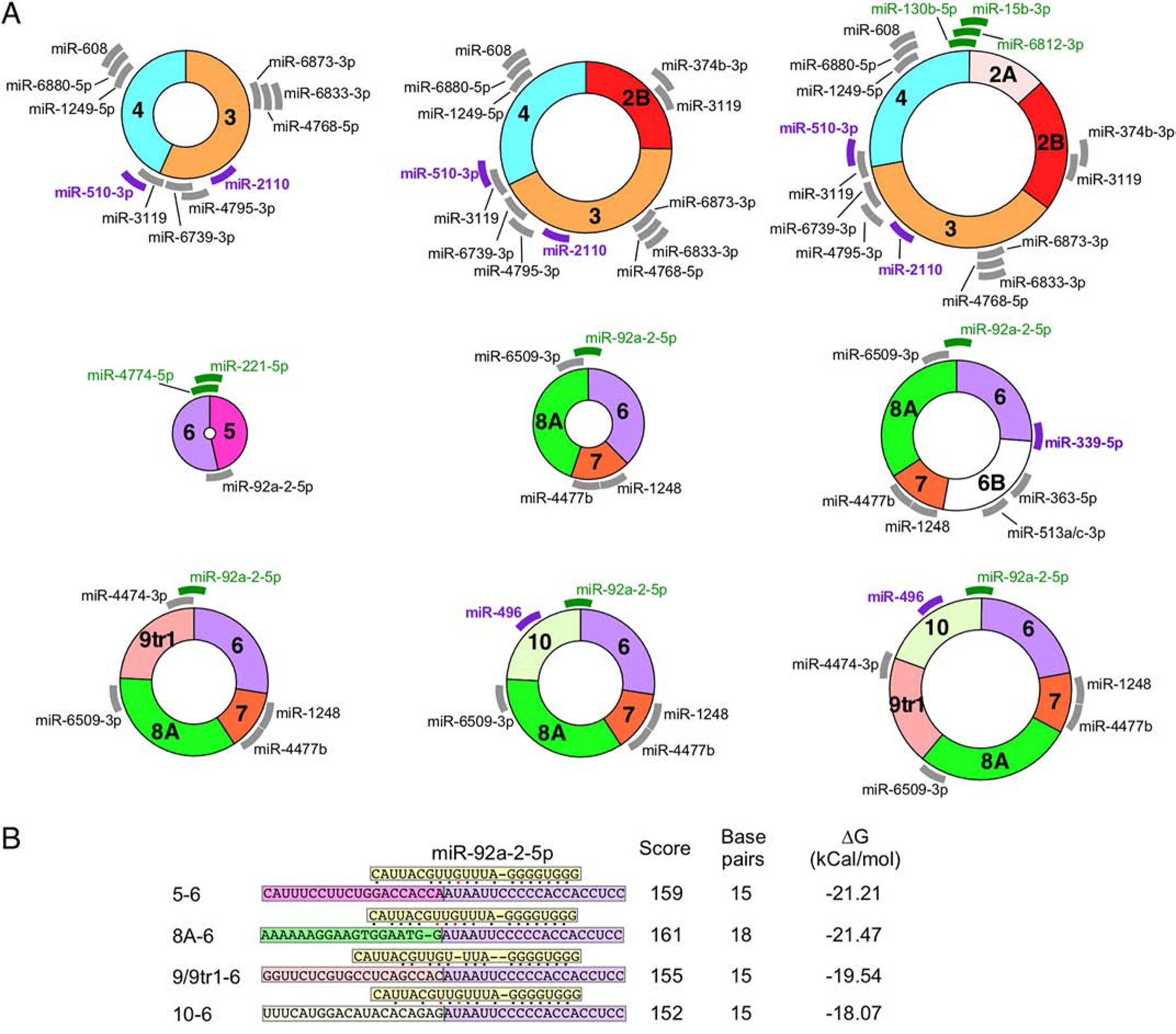
Circular RNAs (circRNAs) belong to a diverse class of stable RNAs expressed in all cell types. Their proposed functions include sponging of microRNAs (miRNAs), sequestration and trafficking of proteins, assembly of multimeric complexes, production of peptides, and regulation of transcription. Backsplicing due to RNA structures formed by an exceptionally high number of Alu repeats lead to the production of a vast repertoire of circRNAs by human Survival Motor Neuron genes, SMN1 and SMN2, that code for SMN, an essential multifunctional protein. Low levels of SMN due to deletion or mutation of SMN1 result in spinal muscular atrophy (SMA), a major genetic disease of infants and children. Mild SMA is also recorded in adult population, expanding the spectrum of the disease. Here we review SMN circRNAs with respect to their biogenesis, sequence features, and potential functions. We also discuss how SMN circRNAs could be exploited for diagnostic and therapeutic purposes.

Keywords: Alu elements; Backsplicing; Spinal muscular atrophy, SMA; Survival motor neuron, SMN; circRNA; microRNA.


Figures

Figure 1.
Figure 1.. Organization of the SMN genes.
A scale depiction of the SMN1 gene (SMN2 has an identical overall structure) and flanking sequences are shown. Exons are depicted by colored shapes. Common exons (≥10% of total SMN RNA) are outlined in black, rare exons (<10% of total SMN RNA) are outlined in red. Repeat sequences as identified by Repeatmasker are depicted by colored arrows. Arrow direction indicates the orientation of the repeat sequence. ISS-N1, a critical splicing regulatory sequence in intron 7, is shown in red. Numbers indicate position within chromosome 5 of the GRCh38 human genome build. Abbreviations: SINEs, short interspersed nuclear elements; LINEs, Long interspersed nuclear elements. Scale and color coding of exons and repeat sequences are given in the boxed region.
Figure 2.
Figure 2.. SMN genes generate a vast repertoire of circRNAs.
The genomic layout of the SMN genes and prominent backsplicing events is given at the top. Exons are shown as colored shapes, introns as broken lines. Exon sizes are given in black below each exon, intron sizes are given in gray above each intron. A cryptic 5′ss in exon 8 is indicated with an arrow. Colored arrows represent backsplicing events. The relative thickness of each arrow represents backsplice site usage frequency; arrow color represents type of circRNA (green for type 1, blue for type 2, and red for type 3). Bottom 4 panels show the total catalog of each type of SMN circRNA. The exon content of each circRNA is indicated graphically. The name of each circRNA is given at the right with total size in parentheses. circRNAs with the highest expression level are boxed.
Figure 3.
Figure 3.. Potential Alu:Alu pairing leading to exon circularization.
(A) Alu-mediated backsplicing of major type 1 circRNAs. Top panel shows the genomic overview of the region from ~2kb upstream of exon 2A to exon 5 and production of C2A-2B-3–4, bottom panel shows the region from exon 2A to exon 5 and production of C2B-3–4 and C3–4. Exons are shown as colored shapes and labeled in bold. Introns are represented by black lines. Repeat elements are represented by colored arrows, the direction of each arrow indicating the orientation of the repeat. Dashed colored lines denote potential pairings between complementary Alu elements. Coloring is arbitrary and is meant to aid in distinguishing between pairings. The circRNA(s) predicted to result from Alu:Alu base pairing is indicated below. Scale and color coding of exons and repeat sequences is given in the boxed region. (B) Alu-mediated backsplicing of C5–6, the major type 2 circRNA. The genomic overview of the region from exon 4 to ~2 kb downstream of exon 6 is given. Coloring and labeling are the same as in (A). (C) Alu-mediated backsplicing of major type 3 circRNAs. The top panel shows the genomic overview of the region from exon 5 to ~2 kb downstream of exon 9 and production of C6–7–8A, C6–6B–7–8A, and C6–7–8A–9/9tr1. The bottom panel shows the genomic overview of the region from exon 5 to ~2kb downstream of exon 10 and production of C6–7–8A–10 and C6–7–8A–9/9tr1–10. Coloring and labeling are the same as in (A).
Figure 4.
Figure 4.. Recognition of exon 8A 5′ss by U1 snRNA.
A diagram of exons 6 through 8 and intervening is given. Exons are shown as colored boxes, introns as lines/broken lines. Splicing events are indicated by dashed arrows. The sequence of exon 8 flanking the 8A 5′ss is given above the exon diagram. Numbering is given relative to the 8A 5′ss. The location of the 5′ss is indicated by an arrow. U1 snRNA is shown above. Base pairs between the 5′ portion of U1 snRNA and the 5′ss are indicated by dots; black dots indicate canonical base pairs and red dots indicate wobble base pairs. The primary splicing product of exon 8A recognition, C6–7–8A, is shown to the right. Of note, other circRNAs including additional exons are also produced downstream of recognition of the 5′ss of exon 8A (See Figures 2 and 3).
Figure 5.
Figure 5.. Predicted miRNA binding to SMN circRNAs.
(A) The 9 SMN circRNAs with the highest predicted expression levels are indicated graphically. Each exon is shown in different colors. miRNA with predicted binding sites located in SMN circRNA are indicated with thick lines with the position representing the predicted binding site. Gray color represents miRNAs of unknown function. Purple color represents miRNAs with identified functions related to SMN biology and/or neurodegeneration. Green color represents miRNAs whose targets are located across the backsplice junction. Thus, these miRNAs bind differentially to circRNA as compared to their linear mRNA counterparts. (B) The predicted binding strength of miR-92a-2–5p targeting different SMN RNAs. miR-92a-2–5p (boxed in yellow) is shown binding to different splice junctions (boxed in exon-specific colors). Base pairs are indicated with dots; black dots indicate canonical base pairs and red dots indicate wobble base pairs. Locations of bulging bases are indicated by dashes. The identity of each junction is indicated at the left. The miRanda score, number of base pairs, and free energy change of binding are given at the right.

References

    1. Mance LG, Mawla I, Shell SM, Cahoon AB, Mitochondrial mRNA fragments are circularized in a human HEK cell line. Mitochondrion. 51 (2020) 1–6. - PubMed
    1. LaRoche-Johnston F F, Monat C, Coulombe S, Cousineau B B, Bacterial group II introns generate genetic diversity by circularization and trans-splicing from a population of intron-invaded mRNAs, PLoS Genet 21(14) (2018) 11:e1007792. - PMC - PubMed
    1. Wang PL, Bao Y, Yee MC, Barrett SP, Hogan GJ, Olsen MN, Dinneny JR, Brown PO, Salzman J, Circular RNA Is Expressed across the Eukaryotic Tree of Life, Plos One 9(3) (2014) 10. - PMC - PubMed
    1. Salzman J, Gawad C, Wang PL, Lacayo N, Brown PO, Circular RNAs Are the Predominant Transcript Isoform from Hundreds of Human Genes in Diverse Cell Types, Plos One 7(2) (2012) 12. - PMC - PubMed
    1. Nigro JM, Cho KR, Fearon ER, Kern SE, Ruppert JM, Oliner JD, Kinzler KW, Vogelstein B, Scrambled exons, Cell 64(3) (1991) 607–13. - PubMed

Publication types

MeSH terms