Review
. 2021 Jan;141(1):124-131.e2.
doi: 10.1016/j.jid.2020.05.093. Epub 2020 Jun 15.

Noninvasive Assessment of Epidermal Genomic Markers of UV Exposure in Skin

Affiliations
Review

Noninvasive Assessment of Epidermal Genomic Markers of UV Exposure in Skin

Elnara Muradova et al. J Invest Dermatol. 2021 Jan.

Abstract

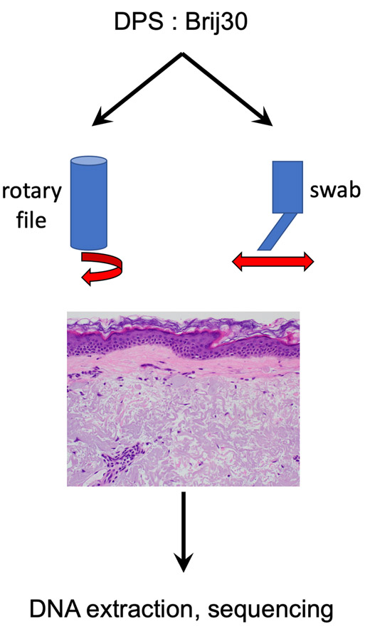
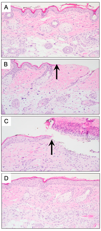
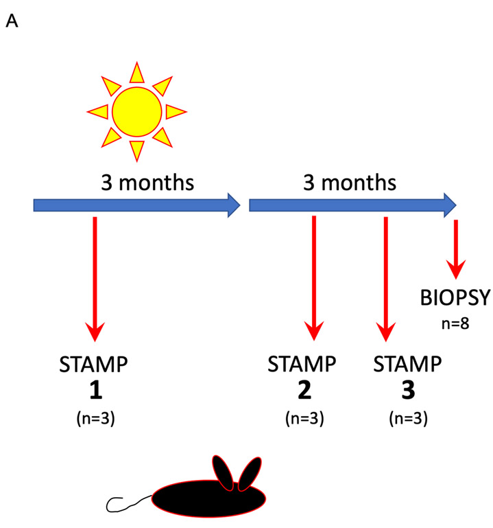
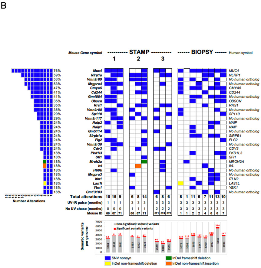
The measurement of UV-induced DNA damage as a dosimeter of exposure and predictor of skin cancer risk has been proposed by multiple groups. Although UV-induced mutations and adducts are present in normal-appearing UV-exposed epidermis, sampling normal nonlesional skin requires noninvasive methods to extract epidermal DNA for analysis. Here, we demonstrate the feasibility of such an approach, termed surfactant-based tissue acquisition for molecular profiling. Sampling in patients was performed using a felt-tip pen soaked in a mixture of surfactants (Brij-30/N-decyl-N,N-dimethyl-3-ammonio-1-propanesulfonate). In mice, we show that the epidermis can be selectively removed without scarring, with complete healing within 2 weeks. We exposed hairless mice to low-dose UV radiation over a period of 3 months and serially sampled them through up to 2 months following the cessation of UV exposure, observing a progressive increase in a UV signature mutational burden. To test whether surfactant-based tissue acquisition for molecular profiling could be applied to human patients, samples were collected from sun-exposed and sun-protected areas, which were then subjected to high-depth targeted exome sequencing. Extensive UV-driven mosaicism and substantially increased mutational loads in sun-exposed versus sun-protected areas were observed, suggesting that genomic measures, as an integrated readout of DNA damage, repair, and clonal expansion, may be informative markers of UV exposure.


Conflict of interest statement

CONFLICTS OF INTEREST

SM and KYT report equity and prior research support from DXB Biosciences / Clearista as well co-inventorship on “Compositions for solubilizing cells and/or tissue”, U.S. Patent number: 9814422. These relationships did not impact funding, study design, or interpretation of results reported.

Figures

Figure 1.
Figure 1.
Surfactant-based Tissue Acquisition for Molecular Profiling (STAMP) The STAMP procedure is accomplished by coupling mechanical energy in the form of rubbing the epidermis with a motorized rotary nail within a chamber full of the surfactant mixture (0.5% w/v each of DPS and Brij-30) or felt swab containing the solution.
Figure 2.
Figure 2.
Skin sampled by STAMP does not scar (A) Full thickness punch biopsy reveals unremarkable skin prior to sampling. (B) Immediately following samples, the epidermis is missing (arrow). (C) At 24 hours post sampling, a fibrinous exudate accumulates. (D) Two weeks following sampling, the epidermis has re-epithelialized, with no evidence of fibrosis or loss of adnexal structures.
Figure 3.
Figure 3.
Dose-dependent accumulation of mutations can be detected using whole-exome sequencing (WES) of skin samples obtained by STAMP in-vivo. (A) The experimental scheme shows the irradiation schema used to produce papillomas and invasive SCC in Hairless mice. In brief, mice were irradiated thrice weekly over 3 months for a total dose of 175 kJ/m2 UVB (broadband). Mice were sampled at 1 month, 2 months, and 5 months using both STAMP and 6 months with standard full-thickness punch biopsy, DNA isolated and processed for whole exome sequencing at 72-140X coverage. (B) Oncoprint showing the burden of mutations in the top 29 most frequently altered genes. Samples shown are from mice sampled by either STAMP (“STAMP”) or punch biopsy (“BIOPSY”). Each column in the center represents one mouse. There are three sets of STAMP samples (each n=3 mice). On the left is the total frequency of mutations observed in these 29 genes in descending order. Several of these genes have no apparent human orthologs or are unlikely to be expressed in skin (e.g. vomeronasal receptors). On the right, the gene names are listed with specific types of variants observed (single nucleotide vs. in-del). The STAMP columns are separated into sets of n=3 mice sampled at 1 month following the start of UV exposure (“1”), 1 month following the cessation of the 3-month course of UV (“2”), and 2 months following the cessation of UV (“3”). The BIOPSY columns represent n=9 full-thickness skin biopsies obtained at 3 months following the cessation of UV. Below, is listed the number of significant variants (red) over total variants (gray) across for each set of samples. The number of significant variants clearly rises over time, achieving an apparent plateau at the 5-6 month time point.
Figure 3.
Figure 3.
Dose-dependent accumulation of mutations can be detected using whole-exome sequencing (WES) of skin samples obtained by STAMP in-vivo. (A) The experimental scheme shows the irradiation schema used to produce papillomas and invasive SCC in Hairless mice. In brief, mice were irradiated thrice weekly over 3 months for a total dose of 175 kJ/m2 UVB (broadband). Mice were sampled at 1 month, 2 months, and 5 months using both STAMP and 6 months with standard full-thickness punch biopsy, DNA isolated and processed for whole exome sequencing at 72-140X coverage. (B) Oncoprint showing the burden of mutations in the top 29 most frequently altered genes. Samples shown are from mice sampled by either STAMP (“STAMP”) or punch biopsy (“BIOPSY”). Each column in the center represents one mouse. There are three sets of STAMP samples (each n=3 mice). On the left is the total frequency of mutations observed in these 29 genes in descending order. Several of these genes have no apparent human orthologs or are unlikely to be expressed in skin (e.g. vomeronasal receptors). On the right, the gene names are listed with specific types of variants observed (single nucleotide vs. in-del). The STAMP columns are separated into sets of n=3 mice sampled at 1 month following the start of UV exposure (“1”), 1 month following the cessation of the 3-month course of UV (“2”), and 2 months following the cessation of UV (“3”). The BIOPSY columns represent n=9 full-thickness skin biopsies obtained at 3 months following the cessation of UV. Below, is listed the number of significant variants (red) over total variants (gray) across for each set of samples. The number of significant variants clearly rises over time, achieving an apparent plateau at the 5-6 month time point.
Figure 4.
Figure 4.
In-vivo STAMP of sun-exposed, at-risk human skin reveals high burden of mutations (A) Two male immunocompromised organ transplant recipients with a history of skin cancer and clinically-evident dermatoheliosis were sampled in both sun-exposed and sun-protected areas and DNA isolated and subjected to targeted exome sequencing to a median depth of 3,000X. (B) Oncoprint of cancer-related genes in sun-exposed vs. sun-protected samples from high-risk organ transplant recipients. The 26 most frequently altered genes in the QIAseq panel are listed in order of decreasing frequency (top to bottom / left to right). Among the most frequently mutated genes are TP53, NOTCH1, MLL2/3, as previously reported. Red boxes denote non-frameshift, non-synonymous variants and black boxes denote frameshift, stopgain, stoploss, or splicing variants. (C) Somatic single-nucleotide variant burdens using both 1% and 2% variant allele cutoffs in sun-exposed and sun-protected samples from high-risk organ transplant recipients.

References

    1. Brash DE. UV signature mutations. Photochem Photobiol 2015;91(1):15–26. - PMC - PubMed
    1. Brash DE, Rudolph JA, Simon JA, Lin A, McKenna GJ, Baden HP, et al. A role for sunlight in skin cancer: UV-induced p53 mutations in squamous cell carcinoma. Proc Natl Acad Sci U S A 1991;88(22):10124–8. - PMC - PubMed
    1. Brash DE, Ziegler A, Jonason AS, Simon JA, Kunala S, Leffell DJ. Sunlight and sunburn in human skin cancer: p53, apoptosis, and tumor promotion. J Investig Dermatol Symp Proc 1996;1(2):136–42. - PubMed
    1. Chitsazzadeh V, Coarfa C, Drummond JA, Nguyen T, Joseph A, Chilukuri S, et al. Cross-species identification of genomic drivers of squamous cell carcinoma development across preneoplastic intermediates. Nat Commun 2016;7:12601. - PMC - PubMed
    1. Diffey BL, Jansen CT, Urbach F, Wulf HC. The standard erythema dose: a new photobiological concept. Photodermatol Photoimmunol Photomed 1997;13(1–2):64–6. - PubMed

Publication types

Substances

LinkOut - more resources