. 2020 Jul:57:102825.
doi: 10.1016/j.ebiom.2020.102825. Epub 2020 Jun 15.

A potent antagonist antibody targeting connexin hemichannels alleviates Clouston syndrome symptoms in mutant mice

Affiliations

A potent antagonist antibody targeting connexin hemichannels alleviates Clouston syndrome symptoms in mutant mice

Yuanyuan Kuang et al. EBioMedicine. 2020 Jul.

Abstract

Background: Numerous currently incurable human diseases have been causally linked to mutations in connexin (Cx) genes. In several instances, pathological mutations generate abnormally active Cx hemichannels, referred to also as "leaky" hemichannels. The goal of this study was to assay the in vivo efficacy of a potent antagonist antibody targeting Cx hemichannels.

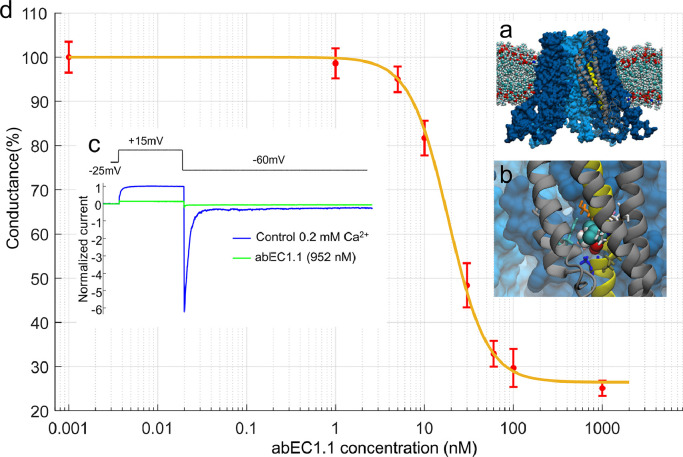
Methods: We employed the antibody to treat Cx30A88V/A88V adult mutant mice, the only available animal model of Clouston syndrome, a rare orphan disease caused by Cx30 p.A88V leaky hemichannels. To gain mechanistic insight into antibody action, we also performed patch clamp recordings, Ca2+ imaging and ATP release assay in vitro.

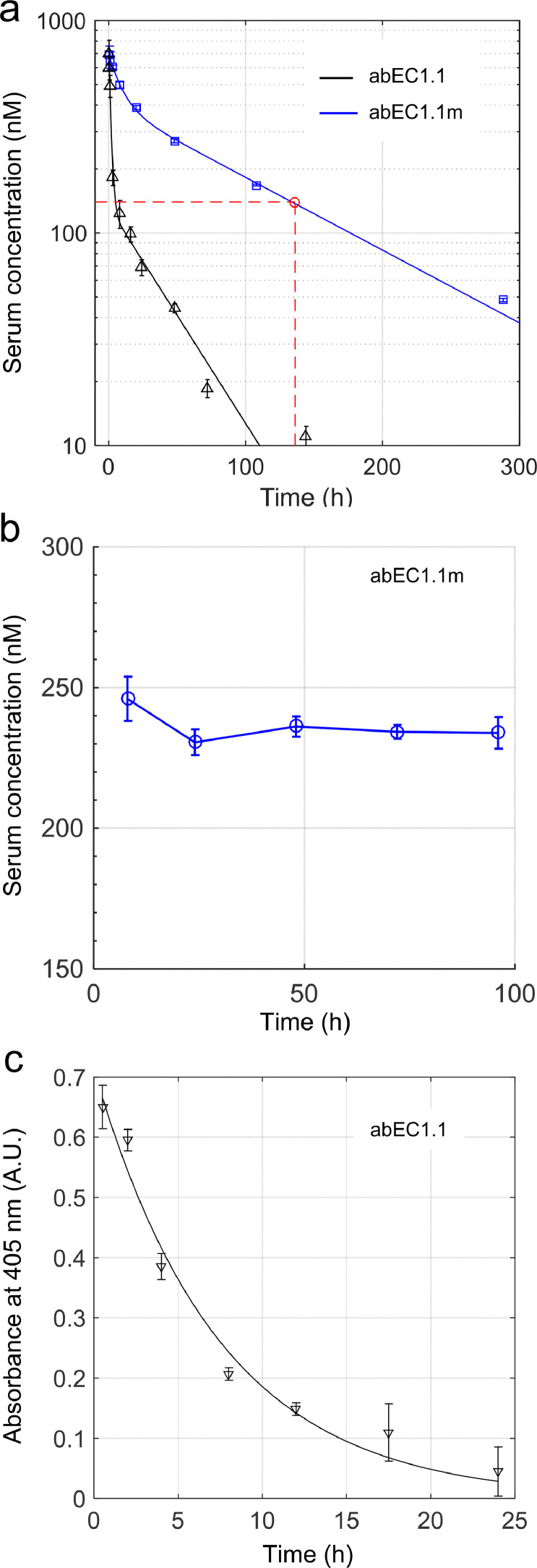
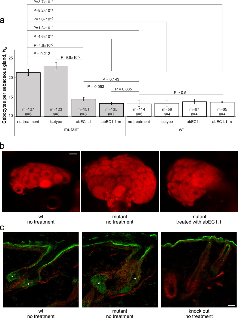
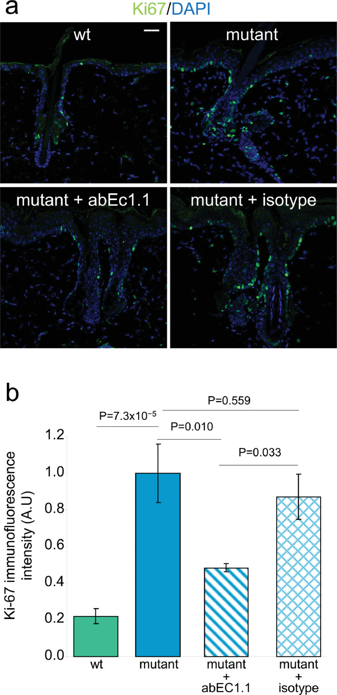
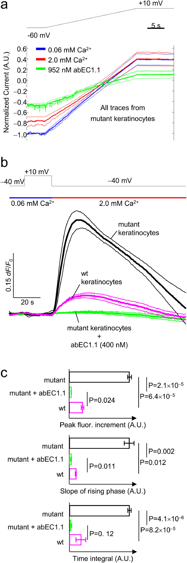
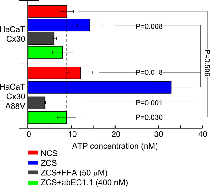
Findings: Two weeks of antibody treatment sufficed to repress cell hyperproliferation in skin and reduce hypertrophic sebaceous glands (SGs) to wild type (wt) levels. These effects were obtained whether mutant mice were treated topically, by application of an antibody cream formulation, or systemically, by intraperitoneal antibody injection. Experiments with mouse primary keratinocytes and HaCaT cells revealed the antibody blocked Ca2+ influx and diminished ATP release through leaky Cx30 p.A88V hemichannels.

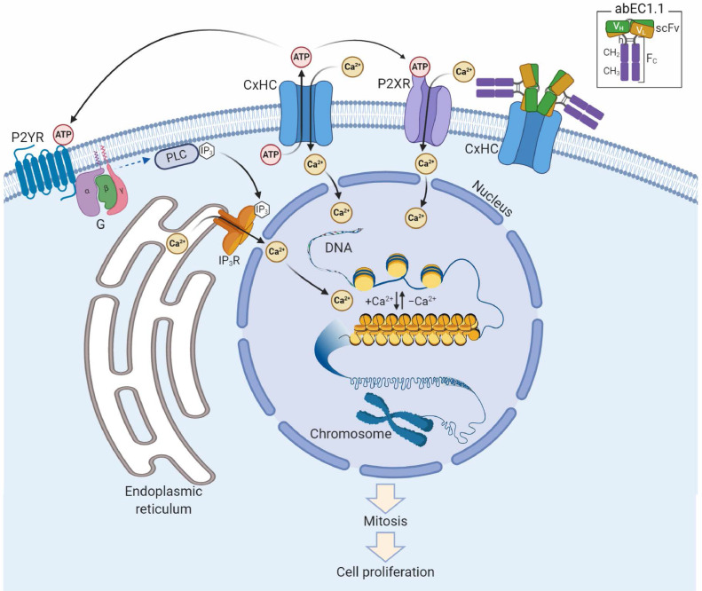
Interpretation: Our results show anti-Cx antibody treatment was effective in vivo and sufficient to counteract the effects of pathological connexin expression in Cx30A88V/A88V mice. In vitro experiments suggest antibodies gained control over leaky hemichannels and contributed to restoring epidermal homeostasis. Therefore, regulating cell physiology by antibodies targeting the extracellular domain of Cxs may enforce an entirely new therapeutic strategy. These findings support the further development of antibodies as drugs to address unmet medical needs for Cx-related diseases. FUND: Fondazione Telethon, GGP19148; University of Padova, SID/BIRD187130; Consiglio Nazionale delle Ricerche, DSB.AD008.370.003\TERABIO-IBCN; National Science Foundation of China, 31770776; Science and Technology Commission of Shanghai Municipality, 16DZ1910200.

Keywords: ATP; Antibody drug discovery; Calcium; Epidermis; Genodermatosis; Sebocytes.


Conflict of interest statement

Declaration of Competing Interest Drs. F. Mammano, G. Yang and F. Zonta report a patent: “Fully human antibody specifically inhibiting connexin 26”, Inventors: Qu Z, Yang G, Mammano F, Zonta F, International application number: PCT/CN2016/109847, pending to ShanghaiTech University; and a patent: “Composition and Methods to treat Ectodermal Dysplasia 2, Clouston Type”, Inventors: Mammano F, Yang G, Zonta F, International Application No.: PCT/CN2019/088689, International Filing Date: 2019-05-28, pending to ShanghaiTech University. All other Authors have nothing to declare.

Figures

Fig 1
Fig. 1
The abEC1.1 antibody inhibits Cx30A88V hemichannel currents with IC50 of 27 nM. a, Molecular model of a Cx30 hemichannel where alanine 88 has been replaced by a valine (A88V mutation). b, Magnified view of the V88 chemical environment. Proximal residues are shown with a licorice representation. c, Representative whole cell currents elicited by shown voltage commands (top, black trace); data were normalized to the mean value of the control response during the application of the +40 mV depolarization step; mean (thick traces) ± s.e.m. (thin traces) for n=5 cells before (blue traces, control) and after application of abEC1.1 at 952 nM for 15 min (green traces) in 0.2 mM Ca2+. d, Percent membrane conductance (mean ± s.e.m. for n ≥ 5 cells), measured with the step protocol shown in panel c and normalized to pre-antibody application levels, vs. abEC1.1 concentration (see Materials and Methods, sections 2.1 and 2.8); the solid line is a least-square fit with a modified Hill equation y = α [1+ (xγ)2]−1 + β, where x is antibody concentration (in nM), α = 73.5, β = (100 − α) = 26.5 and γ = 18.61 nM.
Fig 2
Fig. 2
Antibody levels in serum and skin following single dose administration to C57BL/6N mice. All experiments related to this figure were performed on mice aged 6 to 8 weeks, n = 3 mice per time point (see Materials and Methods, sections 2.4 and 2.5). a, Measurement of abEC1.1 (black) and abEC1.1m (blue) concentration in serum by ELISA vs. time following intravenous bolus injection (100 μl) into the caudal vein at time t = 0 (5 mg of antibody per kg of mouse weight, 5 mg / kg). Each data set was fitted by a double exponential function f (t) = A1 exp(−t /τ1) + A2 exp(−t /τ2) where the coefficients (with 95% confidence bounds) are: A1 = 675 nM (607, 743), τ1 = 1.4 h (1.1, 2.0), A2 = 132 nM (77, 187), τ2 = 43 h (24, 213) for abEC1.1; A1 = 291 nM (230, 352), τ1 = 9.5 h (6.8, 16.1), A2 = 402 nM (338, 466), τ2 = 127 h (101, 171) for abEC1.1m. Intersection of the red dashed lines determines the time (136 h) at which the blood concentration of abEC1.1m falls below 140 nM. b, Measurement of abEC1.1m concentration in serum by ELISA vs. time following intraperitoneal injection at time t = 0 (10 mg of antibody per kg mouse body weight). c, estimate of antibody concentration in skin protein extract by ELISA following topical administration of antibody dispersed in cetomacrogol cream (50 μg / ml); a single application of 100 μl of cream was massaged until completely absorbed in the depilated skin of the mouse back. Data (absorbance at 405 nm; arbitrary units, A.U.) were fitted by an exponential function f (t) = A exp(−t /τ) where the coefficients (with 95% confidence bounds) are: A = 0.71 (0.62, 0.80), τ = 7.2 h (5.9, 10.2), hence t1/2 = ln(2) τ = 5.1 h (4.1, 7.1).
Fig 3
Fig. 3
Effect of antibody treatment on sebaceous glands. All experiments related to this figure were performed on mice aged 6 to 8 weeks (see Materials and Methods, sections 2.2, 2.3, 2.4 and 2.6). a, Counting of Nile red positive cells in sebaceous glands (Ns) of in freshly explanted back skin samples from mutant mice and their wild-type littermates treated topically with abEC1.1 or an inactive isotype antibody, or treated systemically with abEC1.1m; results for non-treated controls are also shown. Legend: n = number of mice; m = number of sebaceous glands; P = p-value (ANOVA). b, Representative multiphoton confocal fluorescence images of Nile red stained sebocytes. Scale bar: 20 μm (see also Supplementary materials, Fig.S2 and Fig. S3). c, Cx30 immunoreactivity in sebaceous glands (asterisks); shown are maximal projection renderings of 10 consecutive confocal optical sections taken at 1 μm intervals; actin filaments were stained with fluorescent phalloidin (red); the green signal is due to a fluorophore-conjugated secondary antibody; in the epidermis and distal end of the hair follicle, the green signal was due to non-specific binding of the secondary antibody, as shown by its persistence in section from Cx30 knock out mice. Scale bar: 20 µm.
Fig 4
Fig. 4
Effect of antibody treatment on the expression of the Ki-67 proliferation marker. All experiments related to this figure were performed on mice aged 6 to 8 weeks (see Materials and Methods, section 2.7). a, Representative transversal sections of mouse dorsal skin showing cells that line the envelope of sebaceous glands labeled with an antibody that binds Ki-67 proteins (green). Shown are maximal projection renderings of nine consecutive confocal optical sections taken at 1.0 μm intervals; scale bar: 30 µm. b, Ki-67 immunoreactivity data (mean ± s.e.m.) obtained by analyzing 7 microscopic fields of view, each field=323 × 323 µm2, from n=3 non-treated mice (wt and mutant littermates) and n=2 treated mice (mutant + abEC1.1 cream, or mutant + isotype antibody cream); P = p-value (ANOVA).
Fig 5
Fig. 5
Effect of acute antibody application on mouse isolated primary keratinocytes. All patch clamp and Ca2+ imaging experiments related to this figure were performed on primary keratinocytes isolated from P2 pups (mutant mice and age-matched wt littermates; see Materials and Methods, sections 2.2, 2.9 and 2.10). a, Hemichannel currents recordings. Voltage ramps (gray trace, top) were applied to the same cell first in 60 µM extracellular Ca2+ concentration ([Ca2+]e) (blue traces), then in 2 mM [Ca2+]e (red traces) or 60 µM [Ca2+]e plus 952 nM [abEC1.1]e (green traces). Currents from each cells were normalized to the level recorded at the −60 mV pre-ramp holding potential in 60 µM [Ca2+]e conditions. Shown are mean values of normalized currents (thick lines) encompassed in 95% confidence intervals (thin lines) for n=3 keratinocytes. b, simultaneous patch clamp and Ca2+ imaging. Top gray trace: voltage clamp protocol; one second before stepping down from +10 mV to −40 mV in 0.06 mM [Ca2+]e (blue horizontal bar) a solution containing 2 mM [Ca2+]e was applied by pressure through a micropipette positioned in the proximity of the patched cells (red horizontal bar). This procedure elicited measurable Fluo-4 signals both in wt keratinocytes (purple traces) and mutant keratinocytes (black traces). In the latter, Fluo-4 signals were abolished by the antibody (green traces). Shown are mean values (thick lines) ± s.e.m. (thin lines) for n=4 wt and n=5 mutant keratinocytes. c, Statistical analysis of the results in panel b. The slope of the rising phase was measured by fitting a straight line to the first 10 s seconds of each fluorescence trace in 2 mM [Ca2+]e conditions at −40 mV; the time integral was measured over the entire trace segment in 2 mM [Ca2+]e conditions at −40 mV. P = p-value (ANOVA).
Fig 6
Fig. 6
Effect of acute antibody application on ATP release in cultured HaCaT cells. All experiments related to this figure were performed in HaCaT cells infected with a lentivirus expressing either human wt Cx30 or human Cx30A88V (see Materials and Methods, section 2.11). NCS, extracellular medium containing a normal 1.8 mM [Ca2+]e; ZCS, extracellular medium containing zero [Ca2+]e; FFA, flufenamic acid. Shown are ATP concentration in the supernatant (mean ± s.e.m.) for n = 3 independent experiments in each of the 4 conditions tested (4 to 12 different cultures in each condition): NCS, ZCS, ZCS + FFA (50 μM) and ZCS + abEC1.1 (400 nM); P=p-values (ANOVA).
Fig 7
Fig. 7
Schematic representation of putative antibody mechanism of action. abEC1.1 (inset) comprises a Cx-binding scFv complex (VH-linker-VL) fused to a fragment constant (Fc) composed of hinge (h), CH2 and CH3 domains of secreted IgG1. Two scFv-Fc homodimers bind the outer vestibule of the connexin hemichannel (CxHC) blocking ATP release and Ca2+ influx [this work and Refs. [35,36]]. The ATP released by open/unblocked CxHCs [this work and Ref. [10]] diffuses in the extracellular milieu and activates G-protein coupled P2Y receptors (P2YR),[29,30] triggering a canonical signal transduction cascade that leads to Ca2+ release from the endoplasmic reticulum via phospholipase C (PLC), PIP2 (not shown) and IP3. At the same time, Ca2+ flows into the cell through ATP-gated P2X ionotropic receptors (P2XR) and open/unblocked CxHCs (this work). Diffusion of Ca2+ in the nucleus promotes transition of chromatin structure from fibrous to globular, chromosome condensation, mitosis and cell proliferation., ,

Comment in

Similar articles

Cited by

References

    1. Srinivas M, Verselis VK, White TW. Human diseases associated with connexin mutations. Biochimica et biophysica acta. 2018;1860(1):192–201. - PMC - PubMed
    1. Lilly E, Sellitto C, Milstone LM, White TW. Connexin channels in congenital skin disorders. Seminars in cell & developmental biology. 2016;50:4–12. - PMC - PubMed
    1. Mammano F. Inner Ear Connexin Channels: Roles in Development and Maintenance of Cochlear Function. Cold Spring Harbor perspectives in medicine. 2019;9(7) - PMC - PubMed
    1. Retamal MA, Reyes EP, Garcia IE, Pinto B, Martinez AD, Gonzalez C. Diseases associated with leaky hemichannels. Frontiers in cellular neuroscience. 2015;9:267. - PMC - PubMed
    1. Garcia IE, Bosen F, Mujica P. From Hyperactive Connexin26 Hemichannels to Impairments in Epidermal Calcium Gradient and Permeability Barrier in the Keratitis-Ichthyosis-Deafness Syndrome. J Invest Dermatol. 2016;136(3):574–583. - PubMed

MeSH terms