. 2020 Jun 30;117(26):15363-15373.
doi: 10.1073/pnas.2002250117. Epub 2020 Jun 17.

Mitochondrial protein interaction landscape of SS-31

Affiliations

Mitochondrial protein interaction landscape of SS-31

Juan D Chavez et al. Proc Natl Acad Sci U S A. .

Abstract

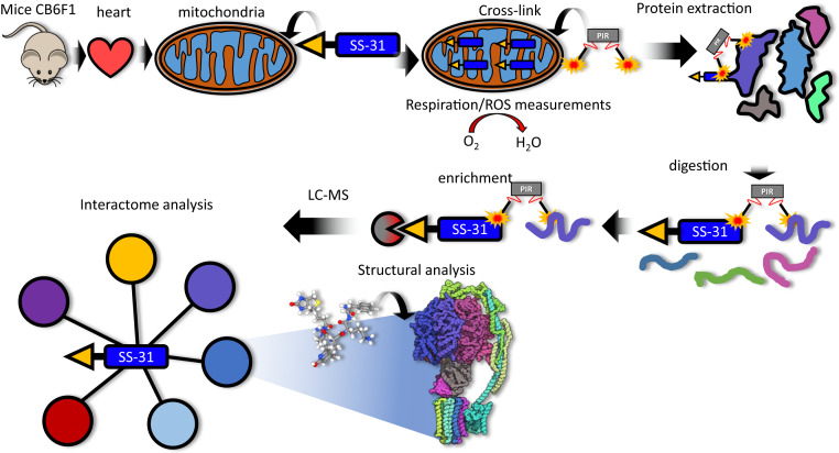
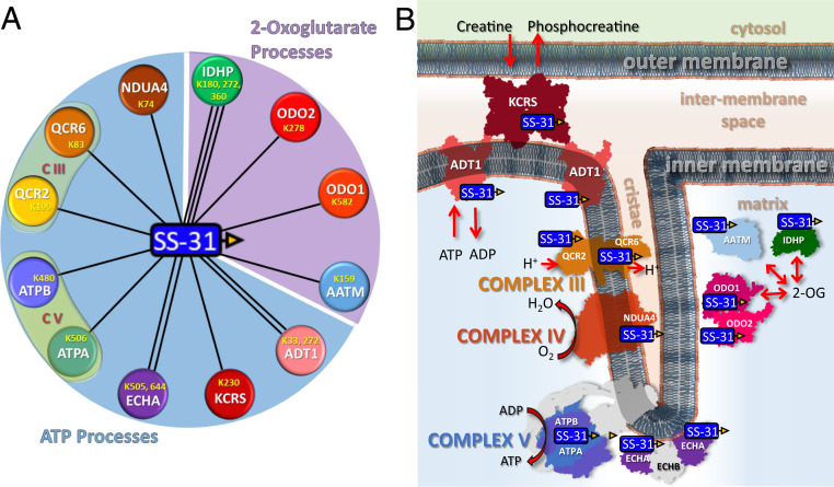
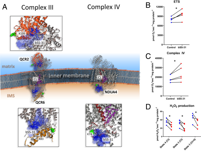
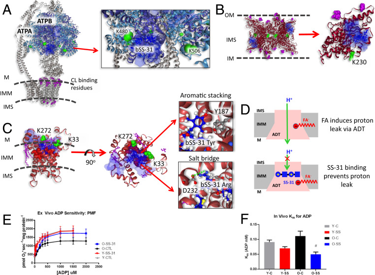
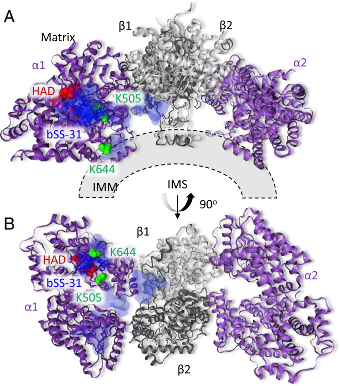
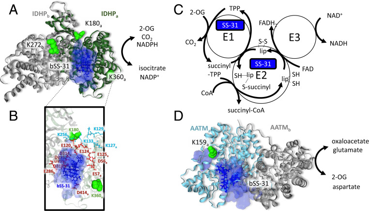
Mitochondrial dysfunction underlies the etiology of a broad spectrum of diseases including heart disease, cancer, neurodegenerative diseases, and the general aging process. Therapeutics that restore healthy mitochondrial function hold promise for treatment of these conditions. The synthetic tetrapeptide, elamipretide (SS-31), improves mitochondrial function, but mechanistic details of its pharmacological effects are unknown. Reportedly, SS-31 primarily interacts with the phospholipid cardiolipin in the inner mitochondrial membrane. Here we utilize chemical cross-linking with mass spectrometry to identify protein interactors of SS-31 in mitochondria. The SS-31-interacting proteins, all known cardiolipin binders, fall into two groups, those involved in ATP production through the oxidative phosphorylation pathway and those involved in 2-oxoglutarate metabolic processes. Residues cross-linked with SS-31 reveal binding regions that in many cases, are proximal to cardiolipin-protein interacting regions. These results offer a glimpse of the protein interaction landscape of SS-31 and provide mechanistic insight relevant to SS-31 mitochondrial therapy.

Keywords: aging; cross-linking; interactome; mitochondria.


Conflict of interest statement

The authors declare no competing interest.

Figures

Fig. 1.
Fig. 1.
Experimental overview. Mice (total of 4), strain CB6F1 (BALB/cBy × C57BL/6), were killed by cervical dislocation. Hearts were excised and processed to isolate mitochondria. Isolated mitochondria were treated with 10 μM biotin SS-31 (bSS-31, blue rectangle with yellow triangle) for 1 h. Oxygen consumption rates and H2O2 production were monitored to evaluate the functional impact of bSS-31 (Fig. 3 BD). Mitochondria were cross-linked with the PIR cross-linker DP-NHP (gray rectangle with orange stars). Protein was extracted using 8 M urea and digested with trypsin. Enrichment for peptides containing bSS-31 was performed with immobilized monomeric avidin. Peptide samples were analyzed by LC-MS to identify the interactome network for bSS-31 and structural analysis was performed using cross-link distance restraint guided molecular docking.
Fig. 2.
Fig. 2.
SS-31 interactome. (A) Nodes represented by colored circles indicate the 12 proteins cross-linked to bSS-31. UniProt identifiers are labeled on the nodes along with the lysine residues that were identified as cross-linked to bSS-31. All 12 proteins are known to interact with cardiolipin (CL, red). The proteins are grouped into two major classes: ATP processes (Upper light blue area) and 2-oxogluterate processes (Lower light purple area). (B) Schematic representation of SS-31 peptide cross-linked proteins within the mitochondria. These proteins include creatine kinase (KCRS), ADP/ATP translocase (ADT1), complex III (CIII) subunits QCR2 and QCR6, complex IV (CIV) subunit NDUA4, complex V (CV) subunits ATPA and ATPB, trifunctional enzyme subunit (ECHA), aspartate aminotransferase (AATM), isocitrate dehydrogenase (IDHP), and 2-oxogluterate dehydrogenase complex subunits ODO1 and ODO2. bSS-31 peptide is represented by a blue rectangle with yellow triangle.
Fig. 3.
Fig. 3.
Structural and functional impact of bSS-31 interaction with OXPHOS complexes CIII and CIV. (A) Structural view of interaction with subunits QCR2 and QCR6 of CIII and NDUA4 of CIV with Inset showing zoomed view of docked structures. Distance constraints of 0 to 35 Å between the α-carbon of K3 of bSS-31 and the α-carbon of cross-linked lysine were used in molecular docking. Proteins are shown in cartoon view. The top 10 molecular docking results for bSS-31 were included as shown in superimposed semitransparent blue surface view with the top scoring docked position of bSS-31 shown in blue ball-and-stick representation. CL and CL binding residues are shown in magenta ball-and-stick and space-filled representation, respectively. Cross-linked lysines are shown in green spheres. (B) Maximum uncoupled respiration in mitochondria isolated from old mouse hearts in the presence of bSS-31 (red) or vehicle control (blue). See SI Appendix, Fig. S3C for data from young mice. (C) Complex IV activity in the presence of bSS-31 (red) or vehicle control (blue). (D) H2O2 production per O2 consumption in mitochondria isolated in the presence of bSS-31 (red) or vehicle control (blue). *P < 0.05 using a paired t test.
Fig. 4.
Fig. 4.
Structural and functional impact of bSS-31 interactions with ATP-producing enzymes. Distance constraints of 0 to 35 Å between the α-carbon of K3 of bSS-31 and the α-carbon of cross-linked lysine residues (green spheres) were used in molecular docking. The top 10 docking results are selected to show the ranges of bSS-31 positions in semitransparent blue surface view with the first docking position displayed in blue ball-and-stick view. CL and CL binding residues are shown in magenta ball-and-stick and space-filled representation, respectively. (A) Structure of bSS-31 interaction with ATP synthase with zoomed Inset. (B) A side view of octameric KCRS (PDB: 1crk) bridging outer and inner membranes of mitochondria is shown in cartoon representation at Left. Shown at Right is monomeric chain A in ribbon view with CL binding residues, K408, R418, and K419, in magenta space-filled balls, and cross-linked residue K230 in green spheres. (C) Structural representation of b-SS31 and ADT interaction in the BKA-locked m-state (PDB: 6gci). Cross-linked lysines K33 and K272 are shown in green spheres and CL is shown in magenta ball-and-stick view. The substrate binding sites, K23, R80, R280, Y187, G183, I184, and S228, are shown in red spheres. Interactive versions of the structures can be viewed at xlinkdb.gs.washington.edu/xlinkdb/BiotinylatedSS31_Bruce.php. Insets show detailed view of salt bridge interaction between D232 residue and Arg residue in bSS-31 and aromatic stacking between Y187 and dimethyl tyrosine in bSS-31. (D) Proposed model of FA-dependent proton leak via ADT (Top) (66), SS-31 binding of ADT possibly prevents proton leak with its two positively charged residues (Bottom). (E) SS-31 treatment increases ADP-stimulated respiration in mitochondria isolated from old mice. (F) SS-31 treatment decreases Km for ADP in vivo in old mice. #P < 0.05 using a paired t test compared to old control. Error bars represent the SEM.
Fig. 5.
Fig. 5.
Structural view of bSS-31 interaction with ECHA. Distance constraints of 0 to 35 Å between the α-carbon of K3 of bSS-31 and the α-carbon of cross-linked lysine residues K644 and K505 of PDB structure 6dv2 were used in molecular docking. The top 10 docking results are selected to show the ranges of bSS-31 positions in semitransparent blue surface view with the first docking position displayed in blue ball-and-stick view. The cross-linked Lys residues on ECHA are shown as green space-filled residues. The HAD active site residues proposed to be important for cardiolipin remodeling (S477, H498, E510, and T548) are shown as red space-filled residues. A displays the side view of the complex, while the view from intermembrane space is shown in B. Interactive versions of the structures can be viewed at xlinkdb.gs.washington.edu/xlinkdb/BiotinylatedSS31_Bruce.php.
Fig. 6.
Fig. 6.
SS-31 interaction with 2-oxoglutarate enzymes. (A) bSS-31 interaction with IDHP (PDB: 5h3f) indicated as a homodimer with the IDHPa chain shown as a green ribbon and the IDHPb chain in gray. Lysine residues cross-linked to bSS-31 are displayed as green space-filled residues and labeled with subscript “a” or “b” to indicate which IDHP monomer they are on. bSS-31 (blue ball-and-stick structure) was docked onto the a-subunit using distance restraints for K180 and K360 from the a-subunit and K272 from the b-subunit. The volume encompassing the positions of bSS-31 from the top 10 docking results is displayed as a semitransparent blue surface. (B) View of the bSS-31 interaction interface highlighting surrounding acidic residues (red) and Lys residues (cyan) which are important in maintaining the groove between the large and small domains of IDHP (84). (C) Schematic representation of the 2-oxoglutarate dehydrogenase complex (2-OGDHC). bSS-31 was identified as cross-linked to ODO1 a component of the E1 of 2-OGDHC and ODO2 a component of the E2 portion of 2-OGDHC. (D) bSS-31 interaction with AATM (PDB: 3pdb) shown as a homodimer with the a-chain in teal and the b-chain in gray. Lysine K159 (green space-filled residue) of the a-chain was used to dock bSS-31 into the structure. The volume encompassing the positions of bSS-31 from the top 10 docking results is displayed as a semitransparent blue surface. Interactive versions of the structures can be viewed at xlinkdb.gs.washington.edu/xlinkdb/BiotinylatedSS31_Bruce.php.

References

    1. Craven L., Alston C. L., Taylor R. W., Turnbull D. M., Recent advances in mitochondrial disease. Annu. Rev. Genomics Hum. Genet. 18, 257–275 (2017). - PubMed
    1. El-Hattab A. W., Zarante A. M., Almannai M., Scaglia F., Therapies for mitochondrial diseases and current clinical trials. Mol. Genet. Metab. 122, 1–9 (2017). - PMC - PubMed
    1. Szeto H. H., Birk A. V., Serendipity and the discovery of novel compounds that restore mitochondrial plasticity. Clin. Pharmacol. Ther. 96, 672–683 (2014). - PMC - PubMed
    1. Szeto H. H., First-in-class cardiolipin-protective compound as a therapeutic agent to restore mitochondrial bioenergetics. Br. J. Pharmacol. 171, 2029–2050 (2014). - PMC - PubMed
    1. Szeto H. H., Stealth peptides target cellular powerhouses to fight rare and common age-related diseases. Protein Pept. Lett. 25, 1108–1123 (2018). - PubMed

Publication types

Substances

LinkOut - more resources