. 2020 Jun 19;368(6497):eabb2751.
doi: 10.1126/science.abb2751.

Meissner corpuscles and their spatially intermingled afferents underlie gentle touch perception

Affiliations

Meissner corpuscles and their spatially intermingled afferents underlie gentle touch perception

Nicole L Neubarth et al. Science. .

Abstract

Meissner corpuscles are mechanosensory end organs that densely occupy mammalian glabrous skin. We generated mice that selectively lacked Meissner corpuscles and found them to be deficient in both perceiving the gentlest detectable forces acting on glabrous skin and fine sensorimotor control. We found that Meissner corpuscles are innervated by two mechanoreceptor subtypes that exhibit distinct responses to tactile stimuli. The anatomical receptive fields of these two mechanoreceptor subtypes homotypically tile glabrous skin in a manner that is offset with respect to one another. Electron microscopic analysis of the two Meissner afferents within the corpuscle supports a model in which the extent of lamellar cell wrappings of mechanoreceptor endings determines their force sensitivity thresholds and kinetic properties.


Conflict of interest statement

Competing Interests

The authors have no competing interests.

Figures

Fig. 1.
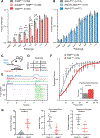
Fig. 1.. Meissner corpuscles and their innervating Aβ-LTMRs are absent in TrkBcKO mice.
A. Digital pad sections from control (upper panels) and TrkBcKO mice (lower panels). Arrows and asterisks indicate typical locations of Meissner corpuscles and Merkel complexes, respectively. Meissner corpuscles and their afferents are labeled with S100 or NFH immunostaining in separate sections, respectively (scale bar = 100 μm). Merkel cells and associated nerve terminals are labeled with TROMA-I and S100, respectively (scale bar = 25 μm). B. Meissner corpuscle and Merkel complex density in glabrous pads of control and TrkBcKO mice. Dots represent individual sections, and black bars represent mean. (79 sections from 3 control mice and 81 sections from 3 TrkBcKO mice; two-tailed Mann-Whitney test, **** p < 0.0001 (U = 580), n.s. = not significant (U = 3163, p = 0.9016)). C. Meissner corpuscle and Merkel complex density in pads of K5Cre; Atoh1flox/+ and Atoh1cKO (K5Cre; Atoh1flox/flox) mice. Dots represent densities from individual sections and black bars represent means. (62 sections from 2 K5Cre; Atoh1flox/+ animals and 53 sections from 2 Atoh1cKO animals; two-tailed Mann-Whitney test, * p = 0.0113 (U = 1193), **** p < 0.0001 (U = 689)). D. In vivo recordings of a slowly adapting (SA, top, blue) and a rapidly adapting (RA, bottom, red) Aβ-LTMR from control mice in response to step indentations applied to glabrous hindpaw pedal pads (force records shown in gray). E. Example recording of an Aβ SA-LTMR from a TrkBcKO mouse in response to the same stimulus as in D. F. Proportion of neurons that are SA, RA, and not responsive (NR) to indentation in control (left, n = 18 cells) and TrkBcKO mice (right, n = 20 cells). χ2 test: χ2 = 8.5, p = 0.004. G. Mean (black bars) and individual (circles) thresholds of Aβ SA-LTMRs in control (left, n = 11 cells) and TrkBcKO mice (right, n = 15 cells). Unpaired t-test: t = −0.81, p = 0.42.
Fig. 2.
Fig. 2.. Meissner corpuscles are necessary for light touch perception and fine sensorimotor control.
A. Fraction (mean ± s.e.m.) of paw withdrawals to von Frey filament applications for TrkBcKO and control mice. RM two-way ANOVA, effect of genotype (F (2,86) = 9.823, p = 0.0001) with post-hoc Tukey’s; p-values represent comparisons between genotypes for each filament: * p < .05, ** p < .01, *** p < .001, **** p < .0001 B. Same as A for Atoh1cKO and control mice. RM two-way ANOVA, no effect of genotype: F (2,34) = 0.8258, p = 0.4465 C. Operant conditioning task design. D. Matrix of possible behavioral outcomes from one trial. E. Operant conditioning task. (Top) Stimulus paradigm, where animals must withhold for at least 3 seconds before initiation of a new trial. On trials with a stimulus (shown), animal receive rewards only on hit trials. (Bottom) Raster plot of licks. Hit trials shown with green ticks, False alarm trials shown with red ticks. F. Psychometric functions for the operant conditioning task for TrkBcKO and control mice. Error bars represent s.e.m. (Inset shows thresholds; unpaired t-test: t = 2.69, p = 0.002). Two-way ANOVA, effect of genotype (F (1,12) = 8.261, p = 0.0140) with post-hoc Sidak’s multiple comparison; p-values represent comparisons between genotypes for each force range: * p < 0.05, ** p < 0.01, *** p < 0.001, **** p < 0.0001. G. Left, the number of sunflower seed touch-taps when the seed is braced against the floor between forepaws during seed peeling. Middle, the number of times the mouse held the seed in an elevated position and used its incisors to bite into the seed while applying downward force (dips) to expose seed kernel. Right, the number of seed rotations as mice adjusted their grasp. Unpaired student’s test, *** p < 0.001 **** p < 0.0001.
Fig. 3.
Fig. 3.. Meissner corpuscles are innervated by two molecularly distinct mechanosensory neuron types.
A. Forelimb pedal pad section of a P20 RetCreER; Rosa26LSL-tdTomato (Ai14) mouse treated with tamoxifen at E10.5-E11.5. Section is immunostained for S100, DsRed, and TO-PRO-3. This experiment was repeated in 3 mice. Scale bar = 25 μm B. Hindlimb digital pad section of a P50 TrkBCreER; Rosa26LSL-tdTomato (Ai14) mouse treated with tamoxifen at E16.5. Section is stained for S100, DsRed, and TO-PRO-3. This experiment was performed in 8 mice with varying dates of tamoxifen administration (E13.5, E16.5, P2, and P6). Scale bar = 50 μm. C. Hindlimb digital pad section of a P50 TrkBCreER; Ai14; RetCFP mouse treated with tamoxifen at P5. Section is immunostained for DsRed, GFP, and NFH. This experiment was repeated in 4 mice with tamoxifen administration at E13.5 or P5. This experiment was repeated in 3 mice. Scale bar = 10 μm. D. Forelimb pedal pad section of a P20 RetCreER; Ai14; Npy2r-GFP mouse treated with tamoxifen at E10.5. Section is stained with anti-DsRed, anti-GFP, and TO-PRO-3. Scale bar = 25 μm. E. Percentage of dermal papillae containing both TrkB+ and Ret+ Meissner afferents in forelimb pads, hindlimb pads, and all pads as measured in TrkBCreER; Ai14; RetCFP mice (2 animals). Only papillae containing a tdTomato+ fiber were included in the quantification to eliminate an effect of Cre-lox efficiency on the interpretation of these measurements.
Fig. 4.
Fig. 4.. Ret+ and TrkB+ Meissner afferents exhibit distinct physiological response properties.
A. Both Meissner afferent subtypes have conduction velocities in the Aβ range as defined by the range of conduction velocities measured from hairy skin Aδ and Aβ LTMRs in the same preparation. The average conduction velocities for TrkB+ and Ret+ Meissner afferents are significantly different (10 afferents per type; mean ± s.e.m.: TrkB = 17.3 ± 0.9 m/s, Ret = 20.9 ± 0.8; Mann-Whitney U Test (U = 26.0, p = 0.008). * p < 0.05. B. The minimal force required to produce an action potential at the onset or offset of a step indentation for TrkB+ and Ret+ Meissner afferents. C. Response of a TrkB+ Meissner Aβ LTMR to a series of step indentations increasing in intensity from 1 to 75 mN. D. Response of a RA Ret+ Meissner afferent to the same stimuli as in C. E. Response of a SA Ret+ Meissner afferent.
Fig. 5.
Fig. 5.. Spatial arrangement of Ret+ and TrkB+ Meissner afferent cutaneous endings.
A. Images of whole-mount, AP-stained digital pads of a TrkBCreER; Rosa26iAP mouse (top) and a RetCreER; Brn3af(AP) mouse (bottom, Z-projection performed in Fiji) depicting pairs of afferents of the same subtype innervating the same glabrous pad. Tamoxifen doses were titrated to achieve sparse labeling of Meissner afferents. Terminals of one neuron are annotated with circles and terminals of the other with triangles. (scale bars = 100 μm) B. Digital pad sections of TrkBCreER; Rosa26LSL-YFP/LSL-tdTomato (Ai3/Ai14) mice treated with tamoxifen at E12.5 and E13.5 (upper panel) and RetCreER; Ai3/Ai14 mice administered tamoxifen at E11.5 and E12.5 (lower panel). Y: fibers express both tdTomato and YFP; R: fibers express tdTomato only; G: fibers express YFP only; G’: YFP+ fibers without terminals in the sections. Sections were stained with anti-DsRed, anti-GFP, and TO-PRO-3. (scale bar = 100 μm) C. Percentage of TrkB+ and Ret+ Meissner afferent pairs innervating the same pad and occupying different spatial territories. D. Quantification of the percentage of dermal papillae and Meissner corpuscles receiving a single TrkB+ fiber (3 animals) and a single Ret+ fiber (6 animals). E. Receptive field surface area (terminal area, left) and number of terminal endings (right) of TrkB+ and Ret+ Meissner afferents measured in TrkBCreER; Rosa26iAP or TrkBCreER; Brn3af(AP) mice (N = 21) and RetCreER; Rosa26iAP or RetCreER; Brn3af(AP) mice (N = 21). Individual measurements and mean values (black bar) are plotted for forelimb digital pads and pedal pads. (two-tailed Welch’s t-test, mean significantly different: * p < .05, ** p < .01; F-test of the equality of variances, variance significantly different: † p < .01).
Fig. 6.
Fig. 6.. TrkB+ Meissner Aβ LTMR endings have more lamellar cell wrappings than Ret+ Meissner mechanoreceptor endings.
A. Image of the paw, with the shaded areas (digit tips) representing regions of high density of Meissner corpuscles used for EM analysis. B. EM images of Meissner corpuscles from a TrkBCreER; AdvillinFlpO; Rosa26DR-Matrix-dAPEX2 mouse treated with tamoxifen at P3 (left) and a RetCreER; AdvillinFlpO; Rosa26DR-Matrix-dAPEX2 mouse treated with tamoxifen at E11.5 and P10 (right). Scale bar = 3 μm. C. High magnification images of labeled endings and associated lamellar cells from (B). Both exhibit openings or areas not directly associated with lamellar cells (black arrowheads). Scale bar = 1 μm. D. Quantification of the number of lamellar cell wrappings around genetically labeled endings of TrkB and Ret endings. Shown are the number of lamellar wrappings for individual axonal profiles (left panel; n = 32 and 47, respectively) and averages of all axonal profiles within individual corpuscles (right panel; n = 12 and 15, respectively). Mann-Whitney U test, **** p < 0.0001. N = 2 animals for each LTMR subtype.

Comment in

  • Getting a grip on touch receptors.
    Marshall K, Patapoutian A. Marshall K, et al. Science. 2020 Jun 19;368(6497):1311-1312. doi: 10.1126/science.abc7610. Science. 2020. PMID: 32554582 No abstract available.

Similar articles

Cited by

References

    1. Wagner R, Meissner G, Über das Vorhandensein bisher unbekannter eigenthümlicher Tastkörperchen (Corpuscula tactus) in den Gefühlswarzchen der menschlichen Haut and über die Endausbreitung sensitiver Nerven. Nachricten von der Georg-August-Universität und der Königl Gesellschaft der Wissenschaften zu Göttingen 2, 17–30 (1852).
    1. González-Martínez T et al., Absence of Meissner corpuscles in the digital pads of mice lacking functional TrkB. Brain Res 1002, 120–128 (2004). - PubMed
    1. González-Martínez T et al., BDNF, but not NT-4, is necessary for normal development of Meissner corpuscles. Neurosci Lett 377, 12–15 (2005). - PubMed
    1. Perez-Pinera P et al., Characterization of sensory deficits in TrkB knockout mice. Neurosci Lett 433, 43–47 (2008). - PubMed
    1. da Silva S et al., Proper formation of whisker barrelettes requires periphery-derived Smad4-dependent TGF-β signaling. Proc Natl Acad Sci U S A 108, 3395–3400 (2011). - PMC - PubMed

Publication types