. 2020 Jun;8(1):e000785.
doi: 10.1136/jitc-2020-000785.

Combined DLL3-targeted bispecific antibody with PD-1 inhibition is efficient to suppress small cell lung cancer growth

Affiliations

Combined DLL3-targeted bispecific antibody with PD-1 inhibition is efficient to suppress small cell lung cancer growth

Xin Chen et al. J Immunother Cancer. 2020 Jun.

Abstract

Background: Small cell lung cancer (SCLC) accounts for 15% of lung cancers, and the primary treatment of this malignancy is chemotherapy and radiotherapy. Delta-like 3 (DLL3) is an attractive target for SCLC immunotherapy since its expression is highly restricted to SCLC with a neglectable appearance on normal adult tissues. In the current study, we aimed to explore the efficacy of DLL3-targeted SCLC immunotherapy via the engagement of T cell.

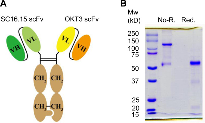
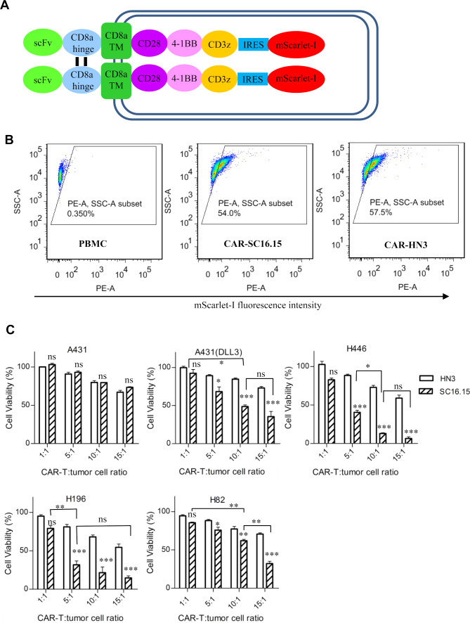
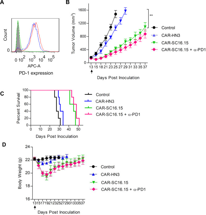
Methods: As a proof of concept, we constructed DLL3-targeted bispecific antibody and chimeric antigen receptor (CAR)-modified T cells. In vitro and in vivo tumor-suppression activity of these treatments alone or in combination with a Program Death-1 (PD-1) inhibitory antibody was evaluated.

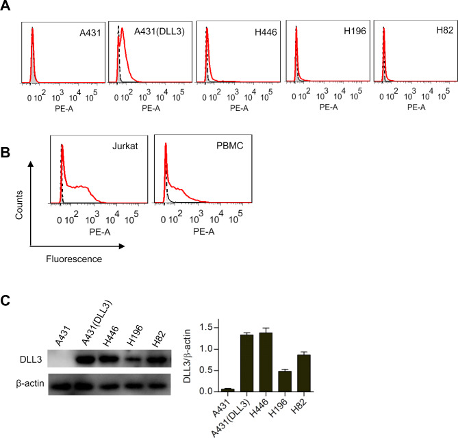
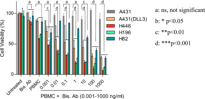
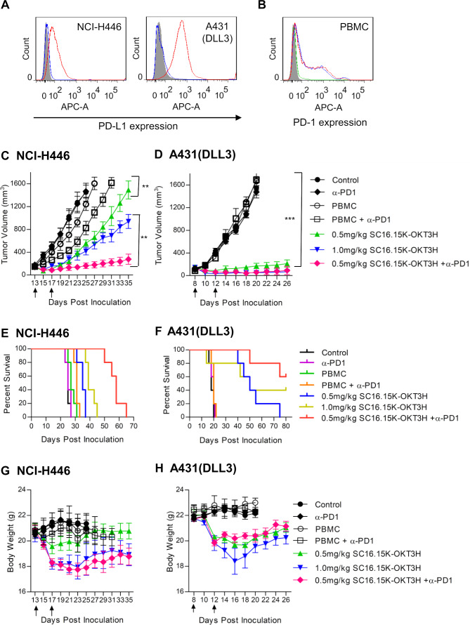
Results: In vitro studies showed that both DLL3 bispecific antibody and CAR-T efficiently killed DLL3-positive cancer cells, including the native SCLC cell lines H446, H196, H82, and the artificial A431 cells that were forcefully overexpressing DLL3. In vivo studies in xenograft mouse models demonstrated that both bispecific antibody and CAR-T suppressed the tumor growth, and combination therapy with PD-1 inhibitory antibody dramatically improved the efficacy of the DLL3 bispecific antibody, but not the CAR-T cells.

Conclusions: Our results demonstrated that DLL3-targeted bispecific antibody plus PD-1 inhibition was effective in controlling SCLC growth.

Keywords: antibodies; biomarkers; immunotherapy; lung neoplasms; neoplasm; tumor.


Conflict of interest statement

Competing interests: None declared.

Figures

Figure 1
Figure 1
Preparation of delta-like 3 (DLL3) bispecific antibody. (A) Schematic diagram of the primary structure of the DLL3 bispecific antibody. The anti-DLL3 scFv (SC16.15) was fused with hFc knob, and the anti-CD3 scFv (OKT3) was fused with hFc hole. (B) SDS-PAGE analysis of the purified bispecific antibody. Two micrograms of protein were loaded for each lane. Non-R., non-reduced condition, showing the dimerized bispecific antibody; Red., 2-mercaptoethanol reduced condition, showing the reduced monomer of the bispecific antibody.
Figure 2
Figure 2
Binding properties of the delta-like 3 (DLL3) bispecific antibody. (A) Flow cytometry analysis of the bispecific antibody binding to different cancer cell lines. Ten micrograms of the bispecific antibody were coincubated with one million of cells. Antibody binding was detected by phycoerythrin-conjugated goat antihuman IgG. Shaded area, secondary antibody staining; dashed lines, isotype control (pooled human IgG) staining; red solid line, bispecific antibody staining. (B) T cell binding analysis of the bispecific antibody. Same experimental settings were used as above mentioned, except that the T cell line Jurkat and peripheral blood mononuclear cells were tested. (C) Western blot analysis of the DLL3 expression in different cancer cell lines. Fifty micrograms of total protein from each cell lysate were run on reduced SDS-PAGE, followed by anti-DLL3 antibody staining. The β-actin was used loading control. A431 (DLL3) was an artificial cell line that was forcefully overexpressing DLL3. The band intensity from each lane was quantified by using ImageJ software, and presented as mean±SEM.
Figure 3
Figure 3
In vitro cell killing assay of the delta-like 3 (DLL3) bispecific antibody. All the tested cell lines were stably transduced to constitutively express a fire fly luciferase reporter gene (ffLuc2). Ten thousand cancer cells were coincubated with two hundred thousand unstimulated peripheral blood mononuclear cells (PBMC) cells for 48 hours in the presence of variable concentrations of the bispecific antibody as indicated. The cell viability was quantified by measuring the intracellular luciferase activity. DLL3-negative A431 was served as a control of the non-specific killing by the bispecific antibody. Statistical comparisons between the A431 control and other cell lines at each bispecific antibody concentration was calculated and labeled on the top of the bar. Data represent mean±SEM.
Figure 4
Figure 4
In vivo efficacy testing of the delta-like 3 (DLL3) bispecific antibody. (A) PD-L1 expression on H446 and A431 (DLL3) cells. One million of cells was stained with PD-L1 rabbit monoclonal antibody, followed by detection with APC-conjugated goat-anti-rabbit IgG. Shaded area, secondary antibody staining; blue curve, isotype control (pooled rabbit IgG) staining; red curve, PD-L1 antibody staining. (B) PD-1 expression on the DLL3 bispecific antibody-stimulated peripheral blood mononuclear cells (PBMC) cells. H446 (red curve) and A431(DLL3) cells (blue curve) were incubated with PBMC for 48 hours in the presence of 100 ng/mL DLL3 bispecific antibody, followed by flow cytometry to analyze PD-1 expression. Shaded area, unstimulated PBMC cell staining; green curve, isotype control (pooled human IgG) staining of H446-stimulated PBMC. (C) Tumor growth curve of native small cell lung cancer cell line NCI-H446. Five million cells were subcutaneously inoculated in each NSG mouse. After the tumor formed and reached a size of 100–200 mm3, treatment was started. Bispecific antibody alone (0.5 mg/kg or 1.0 mg/kg body weight), or in combination of anti-PD-1 antibody (in-house made scFv-hFc format, 5.0 mg/kg) were tested. Ten million unstimulated human PBMC were intraperitoneally given immediately before the first intravenous delivery of the antibody. Arrows indicated the injection time point of the bispecific antibody. The PD-1 antibody was intravenously given once every week since the start of the treatment. Both tumor volume and body weight were measured every two or 3 days. **p<0.01, calculated using a Student’s paired t-test (two-tailed), ***p<0.001. (D) Tumor growth curve of A431 (DLL3), an artificial A431 cell line that was forcefully overexpressing DLL3. ***p<0.001, calculated using a Student’s paired t-test (two tailed). (E) The survival curves of the H446 mouse model treated with the bispecific antibody. (F) The survival curves of the A431 (DLL3) mouse model. (G) Body weight of the H446 mouse model treated with the bispecific antibody. (H) Body weight of the A431 (DLL3) mouse model.
Figure 5
Figure 5
In vitro cell killing assay of the delta-like 3 (DLL3)-targeted chimeric antigen receptor (CAR)-T. (A) Schematic diagram of the CAR structure. Anti-DLL3 scFv (SC16.15) or isotype control (HN3, a human antibody that targets glypican-3, sequence from patent US20140044714) was fused with the following domains, the CD8α hinge, CD8α transmembrane region, CD28 intracellular domain, 4-1BB intracellular domain, CD3ζ intracellular domain, internal ribosome entry site (IRES, from encephalomyocarditis virus), and a red fluorescent protein mScarlet-I. (B) Transduction efficiency analysis of the CAR-T cells, as indicated by the expression of mScarlet-I. Untransduced PBMC was used as negative control. (C) Cell killing measurement of the CAR-T cells. All the cancer cell lines were stably transduced to constitutively express a fire fly luciferase reporter gene (ffLuc2). Ten thousand cancer cells were coincubated with different amount of CAR-T cells for 48 hours. The cell viability was quantified by measuring the intracellular luciferase activity. HN3 was a negative control CAR that targets glypican-3. Data represent mean±SEM. Statistical comparisons between the DLL3 CAR-T (SC16.15) and the control (HN3) at each CAR-T: tumor cell ratio was calculated and labeled on the top of the bar. Comparisons between different DLL3 CAR-T ratios were also calculated. ns, not significant, *p<0.05, ** p<0.01, ***p<0.001.
Figure 6
Figure 6
In vivo efficacy testing of the delta-like 3 (DLL3)-targeted chimeric antigen receptor (CAR)-T cells. (A) PD-1 expression on CAR-T cells. DLL3 CAR-T cells (red curve) and HN3 control CAR-T cells (blue curve) were stained with the PD-1 inhibitory antibody (in-house made scFv-hFc format), followed by APC-conjugated goat-anti-human IgG. Shaded area, peripheral blood mononuclear cells cell staining; green curve, isotype control (pooled human IgG) staining of the DLL3 CAR-T cells. (B) Tumor growth curve of NCI-H446. Five million cells were subcutaneously inoculated in NSG mice. After the tumor formed and reached a size of 100–200 mm3, treatment was started by intraperitoneal delivery of five million CAR-T cells. Arrow indicated the time point of the CAR-T treatment. The anti-PD-1 antibody (in-house made scFv-hFc format, 5.0 mg/kg) was intravenously given once every week since the start of the treatment. Both tumor volume and body weight were measured every 2 or 3 days. **p<0.01, calculated by using a paired Student’s t-test. (C) The survival curves of the treated mice. (D) The body weight of the treated mice.

Similar articles

Cited by

References

    1. Wang S, Zimmermann S, Parikh K, et al. . Current diagnosis and management of small-cell lung cancer. Mayo Clin Proc 2019;94:1599–622. 10.1016/j.mayocp.2019.01.034 - DOI - PubMed
    1. Pavan A, Attili I, Pasello G, et al. . Immunotherapy in small-cell lung cancer: from molecular promises to clinical challenges. J Immunother Cancer 2019;7:205. 10.1186/s40425-019-0690-1 - DOI - PMC - PubMed
    1. Povsic M, Enstone A, Wyn R, et al. . Real-world effectiveness and tolerability of small-cell lung cancer (SCLC) treatments: a systematic literature review (SLR). PLoS One 2019;14:e0219622. 10.1371/journal.pone.0219622 - DOI - PMC - PubMed
    1. Micke P, Faldum A, Metz T, et al. . Staging small cell lung cancer: Veterans Administration Lung Study Group versus International Association for the Study of Lung Cancer--what limits limited disease? Lung Cancer 2002;37:271–6. 10.1016/S0169-5002(02)00072-7 - DOI - PubMed
    1. Waqar SN, Morgensztern D. Treatment advances in small cell lung cancer (SCLC). Pharmacol Ther 2017;180:16–23. 10.1016/j.pharmthera.2017.06.002 - DOI - PubMed

Publication types

MeSH terms

Substances