. 2020 Jun 17;12(548):eaav9760.
doi: 10.1126/scitranslmed.aav9760.

Aptamers against mouse and human tumor-infiltrating myeloid cells as reagents for targeted chemotherapy

Affiliations

Aptamers against mouse and human tumor-infiltrating myeloid cells as reagents for targeted chemotherapy

Adriana De La Fuente et al. Sci Transl Med. .

Abstract

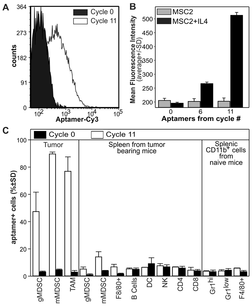
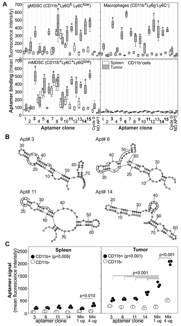
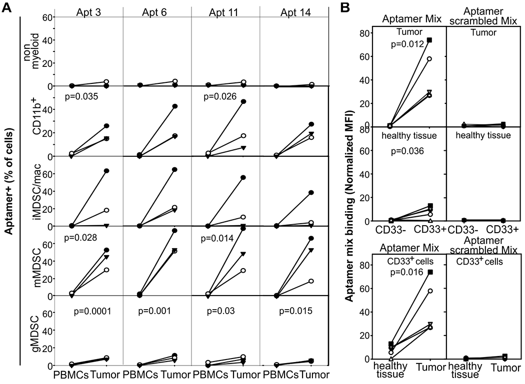
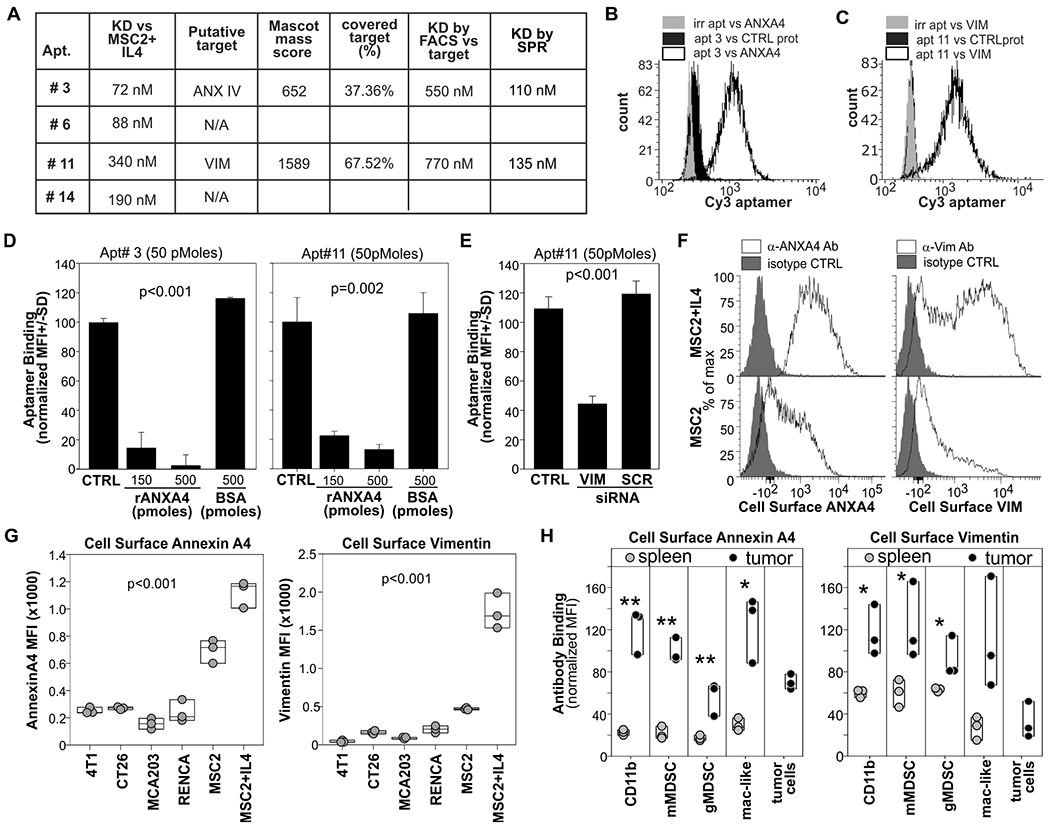
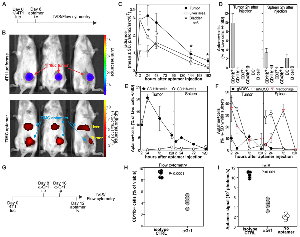
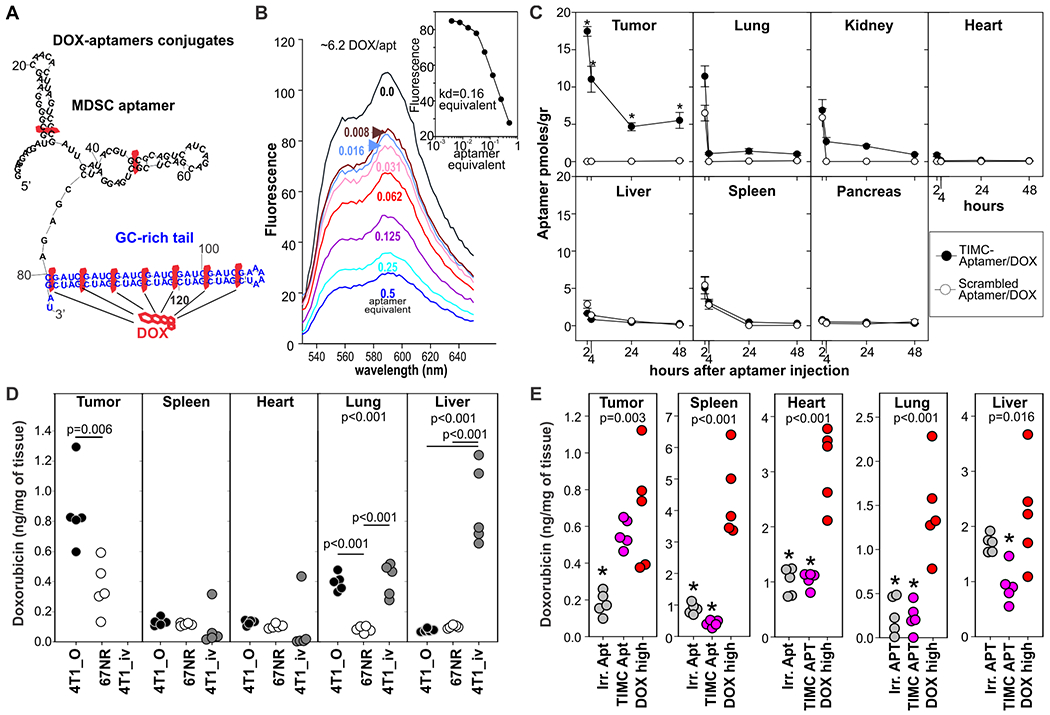
Local delivery of anticancer agents has the potential to maximize treatment efficacy and minimize the acute and long-term systemic toxicities. Here, we used unsupervised systematic evolution of ligands by exponential enrichment to identify four RNA aptamers that specifically recognized mouse and human myeloid cells infiltrating tumors but not their peripheral or circulating counterparts in multiple mouse models and from patients with head and neck squamous cell carcinoma (HNSCC). The use of these aptamers conjugated to doxorubicin enhanced the accumulation and bystander release of the chemotherapeutic drug in both primary and metastatic tumor sites in breast and fibrosarcoma mouse models. In the 4T1 mammary carcinoma model, these doxorubicin-conjugated aptamers outperformed Doxil, the first clinically approved highly optimized nanoparticle for targeted chemotherapy, promoting tumor regression after just three administrations with no detected changes in weight loss or blood chemistry. These RNA aptamers recognized tumor infiltrating myeloid cells in a variety of mouse tumors in vivo and from human HNSCC ex vivo. This work suggests the use of RNA aptamers for the detection of myeloid-derived suppressor cells in humans and for a targeted delivery of chemotherapy to the tumor microenvironment in multiple malignancies.


Conflict of interest statement

Competing interests: A.D., S.Z., J.C, D.V.S. V.B., S.B and P.S. are named as inventors in non-provisional US patent application # 62/815,142 entitled “RNA aptamer and uses thereof” and filed by the University of Miami and regarding the use of RNA aptamer to target myeloid cells. PS is a consultant for Nanovax, LLC.

Figures

Figure 1:
Figure 1:. Selection of polyclonal aptamers specific for tumor infiltrating myeloid cells.
High-throughput Cell-SELEX was performed on MSC2 cells treated with IL4 or left untreated as a proxy of tumor-infiltrating and splenic myeloid cells, respectively. A) FACS analysis of IL4-treated MSC2 stained with the Cy3-labelled RNA aptamers from cycle 0, as control, or cycle 11.B) FACS analysis (N=3) of MSC2 or IL4-treated MSC2 cells with Cy3-labelled polyclonal aptamers from the indicated library. C) Single-cell suspensions from the spleen or tumors of mice bearing CT26 colon carcinomas (0.5 cm in diameter) were stained with a polyclonal aptamer library and counterstained with antibodies against CD11b, Gr1, F4/80, CD11c, CD19, CD49b, CD4, and CD8. Data derived from N=2 experiments with (n=3-5 mice).
Figure 2:
Figure 2:. Identification of monoclonal aptamers specific for tumor-infiltrating myeloid cells.
A) Single-cell suspensions from spleens and tumors of mice pooled from 3 mice bearing 4T1 tumors (0.5 cm in diameter) were labelled with antibodies against CD11b, Ly6C, Ly6G and the 15 Cy3-labelled monoclonal aptamers identified by APTANI. Data derived from n=5 biological replicates and N=2 independent experiments. Aptamers 3, 6, 11, and 14 were chosen for further analysis. * = P<0.001 by one-way ANOVA and post-hoc Holm-Sidak comparisons versus control aptamer. B) Secondary structures of aptamers 3, 6, 11, and 14. Binding motifs identified by APTANI are underlined in black (see table S1). Fluorinated nucleotides are highlighted in gray. C) Specificity of an equimolar mixture of the aptamers for tumor-infiltrating myeloid cells. Pooled single cell suspensions of tumor or spleen from 4T1-bearing mice were labeled with aptamers 3, 6, 11, 14, or equimolar mixtures of each aptamer and analyzed by FACS. Data derived from N=3 independent experiments with pooled spleen and tumors from 5 mice. Data were analyzed by one-way ANOVA and post-hoc Holm-Sidak comparisons.
Figure 3:
Figure 3:. Monoclonal RNA aptamers preferentially recognize human tumor-infiltrating myeloid cells over circulating myeloid cells from patients with recurrent HNSCC.
A) Single-cell suspensions from blood and the tumor of patients (n=3) with recurrent HNSCC were stained with AF647-labelled aptamers, anti-CD33, anti-CD14, anti-CD124 antibodies, and vital dye, and analyzed by flow cytometry. B) Image cytometry: Paraffin-embedded tumor specimens from patients (n=5) with HNSCC undergoing salvage surgery were stained with an equimolar mixture of Cy3-aptamer (TIMC specific or scrambled control), anti-CD33 antibody, and DAPI, and analyzed by cell profiler and FCS-express V6 after gating on the “tumor area” or “healthy tissue area” and either on CD33+ or CD33 cells. Aptamer MFI was normalized on the MFI of the all the cells in the corresponding region of interest. Approximately 106 and 105 cells were analyzed in the “tumor area” or “healthy tissue area”, respectively. Significant paired t-tests are reported.
Figure 4:
Figure 4:. Characteristics of aptamers that preferentially recognize tumor-infiltrating myeloid cells.
A) Affinity of the aptamers for IL4-treated MSC2 cells was determined by flow cytometry. Putative targets (ANXA4 and VIM) were identified via aptamer-based immunoprecipitation, SDS page, and mass spec analysis. Dissociation constants (KDs) against ligands were determined by FACS against recombinant protein conjugated to epoxy beads and by and surface plasmon resonance (SPR) analysis using biotinylated aptamer as ligand and recombinant proteins as analytes. B) Epoxy beads conjugated with ANXA4 or VCAM as control were stained with Cy3-labelled aptamer 3 or Cycle 0 aptamer library and analyzed by FACS. C) Epoxy beads conjugated with rVIM or irrelevant protein were stained with Cy3-labelled aptamer 11 or cycle 0 library and analyzed by FACS. D) ANXA4 and vimentin competitive assay. 5x10⁵ IL4-treated MSC2 were stained with aptamer 3 or aptamer 11 in the presence or in the absence of recombinant ANXA4 or vimentin, respectively. Mean fluorescence intensity (MFI) is reported. E) MCS2 cells were transfected via 4PD nanoparticles with shRNA against vimentin or a scrambled shRNA. 4 days later cells were stained with Cy3-labelled aptamer 11 and DAPI. F) MSC2 or IL4 treated MSC2 were stained withanti-VIM or anti-ANXA4 antibodies or isotype control G) The indicated cell lines were surface-stained with anti-VIM or anti ANXA4 antibodies or isotype control. H) Single-cell suspensions of tumor or spleen of MCA203 tumor-bearing mice with antibodies against CD11b, Ly6g, Ly6c and either vimentin or ANXA4. P= One-way ANOVA p value. *= p<0.05, **= p<0.001. Data derived from at least N=2 independent experiments.
Figure 5:
Figure 5:. Aptamers preferentially target tumor stroma in vivo.
A) Aptamers target tumors in vivo. Mice bearing 4T1-luciferase mammary carcinomas were injected intravenously with an equimolar mixture of biotinylated aptamers 3, 6, 11, and 14 conjugated with AF750 streptavidin. B) Bio-distribution was evaluated by IVIS 2h later. C) Time course analysis or aptamer bio-distribution detected by IVIS. D) Mice (n=5) bearing 4T1 mammary carcinomas were injected i.v. with aptamers 3, 6, 11, and 14 loaded on AF-647 streptavidin. After two hours, indicated organs were harvested and counterstained with antibodies against CD11b, CD19, CD49b, CD11c, and CD3. DC, dendritic cells. E) Mice (n=5) were treated as in (D), and aptamer distribution was evaluated by flow cytometry on CD11b+ and CD11b cells at different time points. F) Mice (n=5) were treated as in (D), and aptamer distribution was evaluated by flow cytometry on gMDSC (CD11b+Ly6g+cells), mMDSC (CD11b+Ly6c+cells), or macrophages (CD11b+F4/80+Ly6gLy6c cells)s. G-I) 4T1-bearing mice were treated i.p with anti-GR1 antibody or rat IgG antibody 8 and 10 days after challenge (G). Two days later, mice were injected i.v. with AF750-conjugated aptamers imaged by IVIS (I), euthanized and tumors analyzed by FACS for the presence of CD11b+cells (H). * = one-way ANOVA p<0.001. Data derived from N=2–3 experiments each with n=3 mice.
Figure 6:
Figure 6:. TIMC-specific aptamers increase doxorubicin concentration at the tumor site.
A) TIMC-specific aptamers were conjugated to DOX by extending the 3’ end with a GC rich tail. B) Fluorescence spectra of DOX solution (1.5 μM) with increasing molar ratios of the TIMC aptamer equimolar mixture (top to bottom: 0, 0.008, 0.016, 0.032, 0.062, 0.125, 0.25, and 0.5 equivalent). Inset: A Hill plot for the aptamer titration (Kd=0.16 equivalent; 6.2 dox molecules/aptamer). C) BALB/c mice (n=5) bearing the orthotopic 4T1 tumors were injected i.v. with DOX (0.35mg/kg) conjugated with TIMC-specific or scrambled aptamers. At the indicated time point mice were euthanized and aptamer biodistribution in different organs was evaluated by qRT-PCR. D) BALB/c mice (n=5) were challenged with orthotopic 4T1 mammary carcinomas (4T1_o; primary tumor: breast, metastases: lung) or intravenously (4T1_iv; metastasis in the lung and in the liver). An additional group of mice was challenged orthotopically with the non-metastatic, 4T1-derived cell line 67NR (only primary tumor). 10 days later, mice were treated i.v. with DOX (0.35 mg/kg) conjugated with TIMC-specific aptamers. DOX bio-distribution was evaluated 24 h later. E) Bio-distribution of free DOX or DOX given via TIMC-specific or scrambled aptamers. Mice (n=5) bearing 4T1 mammary carcinomas (0.5 cm in diameter) in the 3rd mammary gland were given i.v. free doxorubicin (DOX high 3.5 mg/kg), DOX-conjugated TIMC-aptamer (TIMC apt; 0.35mg/kg) or scrambled aptamer (Irr. Apt)-conjugated doxorubicin. 24h later doxorubicin was quantified in tissues by spectrometry after acid alcohol extraction. Data derived from N=1–2 experiments each with n=5 mice *= p<0.05 in multiple pairwise comparison vs dox high group (Holm-Sidak method).
Figure 7:
Figure 7:. TIMC-specific aptamers increase the doxorubicin therapeutic index.
A) Mice bearing 4T1 tumors (~0.5 cm in diameter) in the 3rd mammary gland were treated i.v. with free doxorubicin (3.5 mg/kg or 0.35 mg/kg), Doxil (0.35 mg/kg), or doxorubicin conjugated to a TIMC-specific aptamer mix (0.35 mg/kg). Additional controls included DOX-conjugated scrambled aptamers, unconjugated TIMC-specific aptamers, and untreated mice. Treatment was repeated 2 and 6 days after initial injection. Mice were euthanized when the tumor index reached 1.2 cm2. Log-rank and post-hoc multiple comparison analysis (Holm-Sidak method) is reported. Individual tumor growth curves are shown in fig. S6. B) C57Bl/6 mice were challenged with MCA203 fibrosarcoma. When tumors reached ~0.5cm in diameter, mice were treated with vehicle, high doses of free DOX (3.5 mg/kg), or low doses of DOX (0.35 mg/kg) conjugated either to TIMC-specific or scrambled aptamer. C, D) Weight loss as measure of toxicity is reported for mice bearing the 4T1 (C) and MCA203 (D) tumors. E) BALB/c mice were challenged with the 4T1wt breast cancer or with a DOX-resistant variant of 4T1. When tumors reached 0.5 cm in diameter, mice were treated with TIMC-specific aptamers or left untreated. Survival was monitored. Data are cumulative of 2-3 independent experiments.

Similar articles

Cited by

References

    1. Carvalho C, Santos RX, Cardoso S, Correia S, Oliveira PJ, Santos MS, Moreira PI, Doxorubicin: the good, the bad and the ugly effect. Current medicinal chemistry 16, 3267–3285 (2009). - PubMed
    1. Kydd J, Jadia R, Velpurisiva P, Gad A, Paliwal S, Rai P, Targeting Strategies for the Combination Treatment of Cancer Using Drug Delivery Systems. Pharmaceutics 9, (2017). - PMC - PubMed
    1. Kinnear C, Moore TL, Rodriguez-Lorenzo L, Rothen-Rutishauser B, Petri-Fink A, Form Follows Function: Nanoparticle Shape and Its Implications for Nanomedicine. Chemical reviews 117, 11476–11521 (2017). - PubMed
    1. Bahrami B, Hojjat-Farsangi M, Mohammadi H, Anvari E, Ghalamfarsa G, Yousefi M, Jadidi-Niaragh F, Nanoparticles and targeted drug delivery in cancer therapy. Immunology letters 190, 64–83 (2017). - PubMed
    1. Tiet P, Berlin JM, Exploiting homing abilities of cell carriers: Targeted delivery of nanoparticles for cancer therapy. Biochemical pharmacology, (2017). - PMC - PubMed

Publication types