. 2020 Jun 17;5(3):e00313-20.
doi: 10.1128/mSphere.00313-20.

The Functional Characterization of TcMyoF Implicates a Family of Cytostome-Cytopharynx Targeted Myosins as Integral to the Endocytic Machinery of Trypanosoma cruzi

Affiliations

The Functional Characterization of TcMyoF Implicates a Family of Cytostome-Cytopharynx Targeted Myosins as Integral to the Endocytic Machinery of Trypanosoma cruzi

Nathan Michael Chasen et al. mSphere. .

Abstract

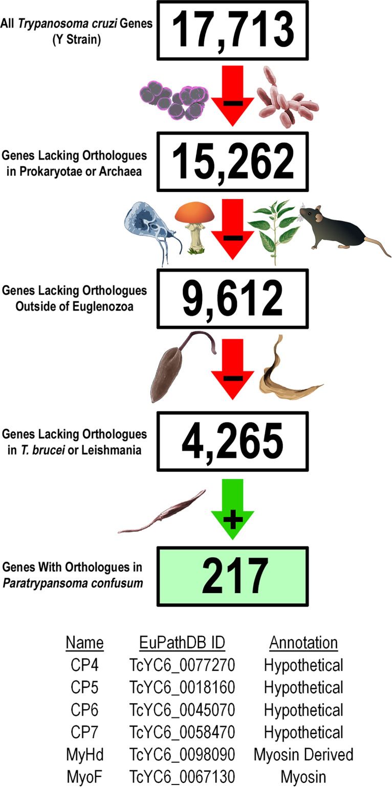
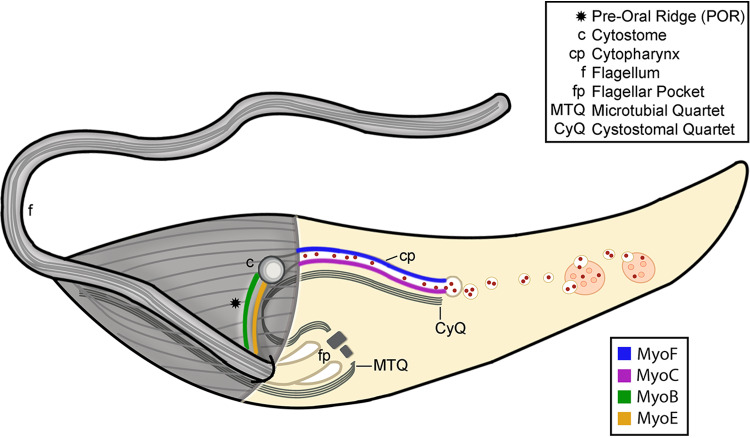
Of the pathogenic trypanosomatids, Trypanosoma cruzi alone retains an ancient feeding apparatus known as the cytostome-cytopharynx complex (SPC) that it uses as its primary mode of endocytosis in a manner akin to its free-living kinetoplastid relatives who capture and eat bacterial prey via this endocytic organelle. In a recent report, we began the process of dissecting how this organelle functions by identifying the first SPC-specific proteins in T. cruzi Here, we continued these studies and report on the identification of the first enzymatic component of the SPC, a previously identified orphan myosin motor (MyoF) specifically targeted to the SPC. We overexpressed MyoF as a dominant-negative mutant, resulting in parasites that, although viable, were completely deficient in measurable endocytosis in vitro To our surprise, however, a full deletion of MyoF demonstrated only a decrease in the overall rate of endocytosis, potentially indicative of redundant myosin motors at work. Thereupon, we identified three additional orphan myosin motors, two of which (MyoB and MyoE) were targeted to the preoral ridge region adjacent to the cytostome entrance and another (MyoC) which was targeted to the cytopharynx tubular structure similar to that of MyoF. Additionally, we show that the C-terminal tails of each myosin are sufficient for targeting a fluorescent reporter to SPC subregions. This work highlights a potential mechanism used by the SPC to drive the inward flow of material for digestion and unveils a new level of overlapping complexity in this system with four distinct myosin isoforms targeted to this feeding structure.IMPORTANCE The parasite Trypanosoma cruzi is the etiological agent of Chagas disease and chronically infects upwards of 7 million people in the Americas. Current diagnostics and treatments remain grossly inadequate due, in part, to our general lack of understanding of this parasite's basic biology. One aspect that has resisted detailed scrutiny is the mechanism employed by this parasite to extract nutrient resources from the radically different environments that it encounters as it transitions between its invertebrate and mammalian hosts. These parasites engulf food via a tubular invagination of its membrane, a strategy used by many protozoan species, but how this structure is formed or functions mechanistically remains a complete mystery. The significance of our research is in the identification of the mechanistic underpinnings of this feeding organelle that may bring to light new potential therapeutic targets to impede parasite feeding and thus halt the spread of this deadly human pathogen.

Keywords: SPC; Trypanosoma cruzi; cytopharynx; cytostome; endocytosis; myosin; reservosome.


Figures

FIG 1
FIG 1
(Top panel) Bioinformatic identification of potential SPC proteins. The list of 17,713 annotated T. cruzi genes from the Y strain genome assembly was narrowed down to 9,612 genes that do not have orthologues outside the Euglena genus. This list was further trimmed to 4,265 genes by eliminating all genes that have orthologues in Leishmania and T. brucei which lack the SPC. The final filter eliminated genes that did not have orthologues in the monoxenous kinetoplastid parasite Paratrypanosoma confusum, which has an SPC, resulting in a list of 217 potential SPC genes. (Bottom panel) List of seven proteins that have been shown to localize to the SPC. ID, identifier.
FIG 2
FIG 2
Identification and localization of six SPC proteins. (A) CP4 localizes to the SPC, potentially on the CyQ and MtQ (arrow) microtubule root fibers and the basal bodies associated with the flagellum (arrowhead). (B to D) CP5 (B), CP6 (C), and CP7 (D) localize to the POR of the SPC labeled by the lectin concanavalin A (purple arrows). CP5 also shows basal body localization (arrowhead). (E and F) Two proteins annotated to contain myosin motor domains (MyoF [E] and MyHd [F]) localize to the cytopharynx of the SPC. Nuclei and kinetoplasts in all fluorescent images were stained with DAPI (4′,6-diamidino-2-phenylindole) (blue). Scale bars, 2 μM.
FIG 3
FIG 3
Dominant-negative MyoF mutants are defective in SPC endocytosis. (A) Schematic of the MyoF gene showing the annotated myosin and SCP1 coiled-coil domains. (B) I-TASSER structure prediction of MyoF (colors) aligned with a known myosin crystal structure (gray) showing ATP binding P-loop region (green) and switch regions (magenta). A conserved lysine residue that is essential for P-loop function is shown in red (K198, arrow). (C) Sequence of the ATP binding P-loop region showing the K198 amino acid residue (red) that was mutated to a glutamine (K198Q) to generate a rigor mutant of MyoF (MyoF-K198Q) for dominant-negative expression. (D) Immunoblot probed with anti-Ty antibody showing similar levels of MyoF and MyoF-K198Q in the chosen subclones. LC, loading control. (E and F) Overexpressed MyoF (E) and MyoF-K198Q (F) fused to mNeon exhibited the same localization in epimastigotes. (G and H) Flow cytometry of epimastigotes after fluorescent BSA feeding shows that the actin inhibitor cytochalasin D prevents SPC endocytosis. (I and J) Flow cytometry of epimastigotes after fluorescent BSA feeding shows a dominant-negative endocytosis defect in the MyoF-K198Q mutants. G and I, counts; H and J, percent positive. Nuclei and kinetoplasts in all fluorescent images were stained with DAPI (blue). Scale bars, 2 μM.
FIG 4
FIG 4
MyoF deletion mutants exhibit a reduction in the rate of SPC endocytosis. (A) Scheme for CRISPR/Cas9 gene replacement and complementation strategy used to generate ΔMyoF deletion and complemented (Δ::MyoF-Ty) mutants. GAPDH, glyceraldehyde-3-phosphate dehydrogenase. (B) PCR amplification of the genomic locus showing replacement of both parental loci (high-molecular-weight [MW] band) with the blasticidin resistance gene (low-MW band) in a subclone of the ΔMyoF mutant. PCR of a nonsubcloned population of complemented parasites shows insertion of the MyoF-Ty-Hygro repair template into the MyoF locus. (C) Anti-Ty immunoblot of lysates from parental, ΔMyoF, and (Δ::MyoF-Ty) epimastigotes showing expression of MyoF-Ty in the complemented parasites. (D) Immunofluorescence assays of a (Δ::MyoF-Ty) epimastigote showing expression and proper SPC localization of MyoF-Ty. (E) Flow cytometry analysis of epimastigotes fed fluorescent BSA showing a reduced rate of feeding in ΔMyoF mutants, a phenotype that is partially rescued in the complemented (Δ::MyoF-Ty) mutants. (F) Quantification of the feeding rate results represented by reduced mean fluorescence of endocytosing epimastigotes. A dramatic reduction in the feeding rate was seen in the ΔMyoF mutants, which was partially rescued by complementation (Δ::MyoF-Ty). (G) A minor but statistically significant percentage of the total number of ΔMyoF and (Δ::MyoF-Ty) epimastigotes showed reduced levels of endocytosed fluorescent BSA during the assay.
FIG 5
FIG 5
Three additional T. cruzi myosins localize to the SPC. (A) Overexpression and superresolution structured illumination (SR-SIM) microscopy of MyoC-mNeon revealed cytopharynx localization, suggesting that MyoC may provide redundancy to the cytopharynx endocytic machinery. (B and C) MyoB (B) and MyoE (C) both localize to the preoral ridge of the SPC (purple arrows). Nuclei and kinetoplasts in all fluorescent images were stained with DAPI (blue). Scale bars, 2 μM.
FIG 6
FIG 6
The C-terminal tail regions of myosin F, C, B, and E are sufficient for SPC targeting. (A) Schematic of myosin genes and the regions (green and red lines) that were fused to the C terminus of mNeon for localization. The green-colored regions were sufficient for localization to the SPC when fused to mNeon, whereas the red-colored regions were not. (B, C, and D) The MyoF-Ct1 (B) and MyoF-Ct3 (D) regions were sufficient to target mNeon to the SPC cytopharynx, although MyoF-Ct3 had a notably less defined localization. MyoF-Ct2 (C) was not sufficient for SPC targeting but did demonstrate localization adjacent to the kinetoplast (arrowhead). (E and F) MyoC-Ct2 (F) was sufficient for targeting mNeon to the SPC cytopharynx, whereas MyoC-Ct1 (E) was not. (G and H) The C-terminal regions of MyoB (G) and MyoE (H) were both sufficient to target mNeon to the SPC preoral ridge. Nuclei and kinetoplasts in all fluorescent images were stained with DAPI (blue). Scale bars, 2 μM.
FIG 7
FIG 7
A family of orphan myosins target to distinct regions of the SPC. A cartoon schematic of a T. cruzi epimastigote is shown, highlighting the internal and external structures of the cytostome-cytopharynx complex. The microtubules of the axoneme, cytoskeleton, and root fibers are depicted in gray. The individual components of the SPC and parasite body are referenced in the associated key. Different colored lines denote the observed locations of the four orphan myosins targeted to distinct subregions of the cytostome-cytopharynx complex.

Similar articles

Cited by

References

    1. Weekly Epidemiological Record. 2015. Chagas disease in Latin America: an epidemiological update based on 2010 estimates. Wkly Epidemiol Rec 90:33–43. - PubMed
    1. Pérez-Molina JA, Molina I. 2018. Chagas disease. Lancet 391:82–94. doi:10.1016/S0140-6736(17)31612-4. - DOI - PubMed
    1. Groom ZC, Protopapas AD, Zochios V. 2017. Tropical diseases of the myocardium: a review. Int J Gen Med 10:101–111. doi:10.2147/IJGM.S130828. - DOI - PMC - PubMed
    1. Camandaroba EL, Reis EA, Goncalves MS, Reis MG, Andrade SG. 2003. Trypanosoma cruzi: susceptibility to chemotherapy with benznidazole of clones isolated from the highly resistant Colombian strain. Rev Soc Bras Med Trop 36:201–209. doi:10.1590/s0037-86822003000200002. - DOI - PubMed
    1. Mejía-Jaramillo AM, Fernández GJ, Montilla M, Nicholls RS, Triana-Chávez O. 2012. Trypanosoma cruzi strains resistant to benznidazole occurring in Colombia. Biomedica 32:196–205. (In Spanish.) - PubMed