

The radiological diagnosis of bronchiectasis: what's in a name?
- PMID: 32554759
- PMCID: PMC9489191
- DOI: 10.1183/16000617.0120-2019
The radiological diagnosis of bronchiectasis: what's in a name?
Abstract
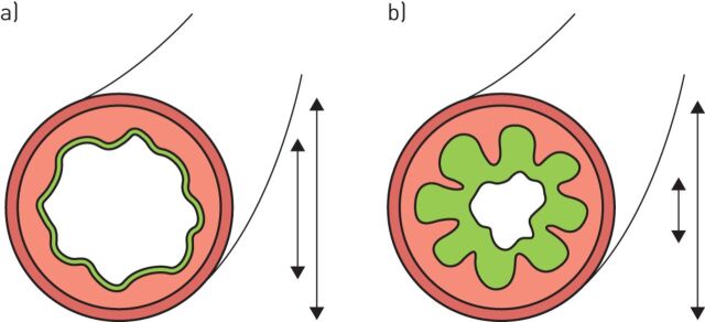
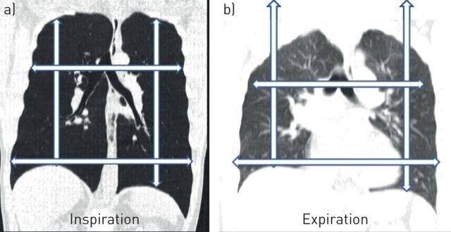
Diagnosis of bronchiectasis is usually made using chest computed tomography (CT) scan, the current gold standard method. A bronchiectatic airway can show abnormal widening and thickening of its airway wall. In addition, it can show an irregular wall and lack of tapering, and/or can be visible in the periphery of the lung. Its diagnosis is still largely expert based. More recently, it has become clear that airway dimensions on CT and therefore the diagnosis of bronchiectasis are highly dependent on lung volume. Hence, control of lung volume is required during CT acquisition to standardise the evaluation of airways. Automated image analysis systems are in development for the objective analysis of airway dimensions and for the diagnosis of bronchiectasis. To use these systems, clear and objective definitions for the diagnosis of bronchiectasis are needed. Furthermore, the use of these systems requires standardisation of CT protocols and of lung volume during chest CT acquisition. In addition, sex- and age-specific reference values are needed for image analysis outcome parameters. This review focusses on today's issues relating to the radiological diagnosis of bronchiectasis using state-of-the-art CT imaging techniques.
Copyright ©ERS 2020.
Conflict of interest statement
Conflict of interest: H.A.W.M. Tiddens has received other funding from Roche and Novartis for symposium, lecture fees and advisory boards. He has received grants from CFF, Vertex, Gilead, Chiesi and Vectura outside the submitted work. He also has a patent pending for the PRAGMA-CF scoring system. He is head of the Erasmus MC core laboratory Lung Analysis which is a not-for-profit core image analysis laboratory. The financial aspects of the laboratory are handled by the department of Radiology and by the Sophia Research BV. FLUIDDA has developed computational fluid dynamic modelling based on chest CTs obtained from Erasmus MC-Sophia for which royalties are received by Sophia Research BV. Conflict of interest: J.J. Meerburg has nothing to disclose. Conflict of interest: M.M. van der Eerden has nothing to disclose. Conflict of interest: P. Ciet has nothing to disclose.
Figures
References
-
- Boaventura R, Shoemark A, Chalmers JD. Pathophysiology. In: Chalmers JD, Polverino E, Aliberti S, eds. Bronchiectasis (ERS Monograph). Sheffield, European Respiratory Society, 2018; pp. 8–28.