Observational Study
. 2020 Jun 17;12(12):12324-12341.
doi: 10.18632/aging.103441. Epub 2020 Jun 17.

Ovarian aging increases small extracellular vesicle CD81+ release in human follicular fluid and influences miRNA profiles

Affiliations
Observational Study

Ovarian aging increases small extracellular vesicle CD81+ release in human follicular fluid and influences miRNA profiles

Rosalia Battaglia et al. Aging (Albany NY). .

Abstract

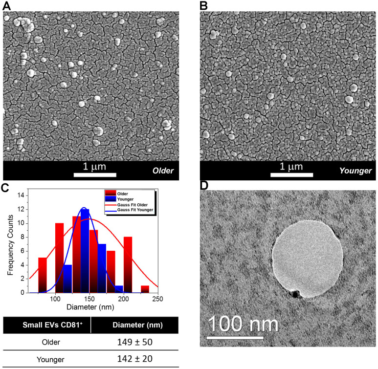
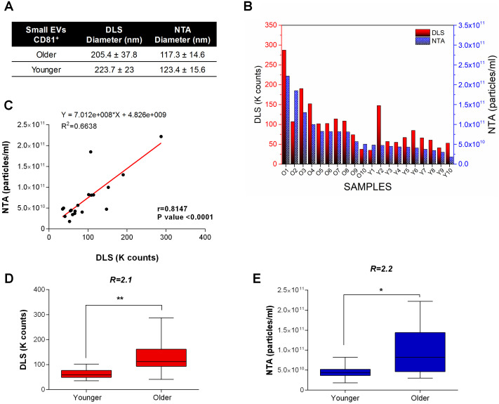
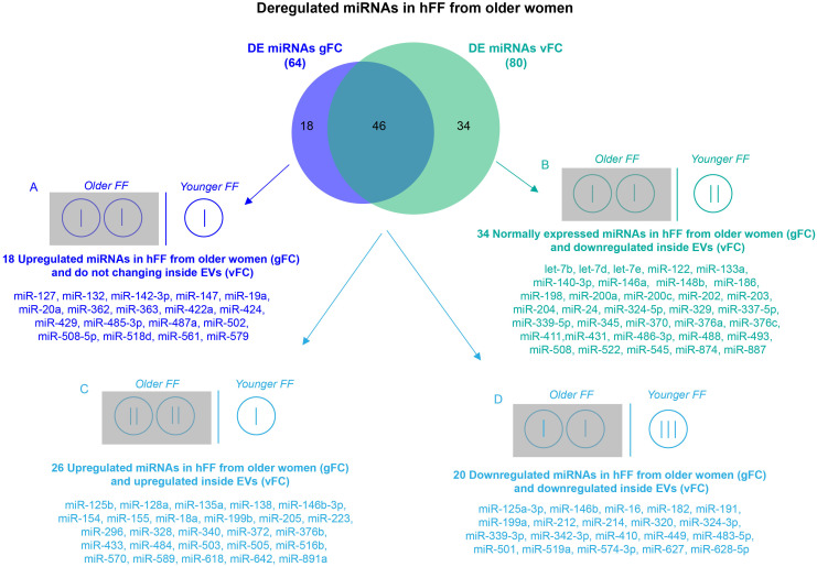
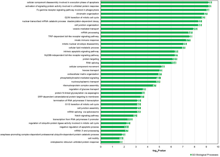
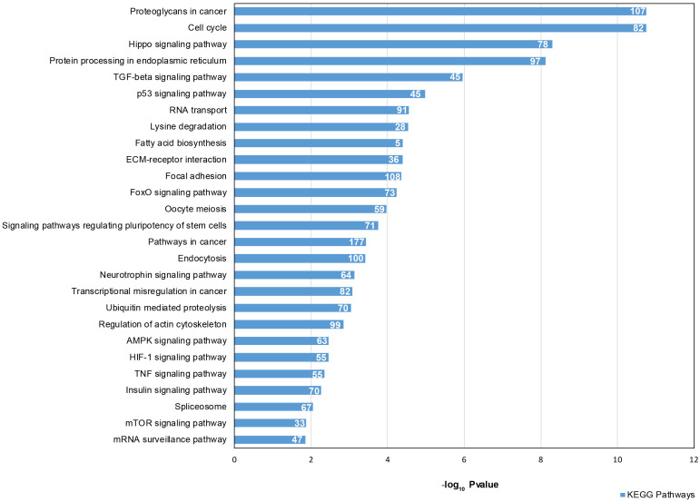
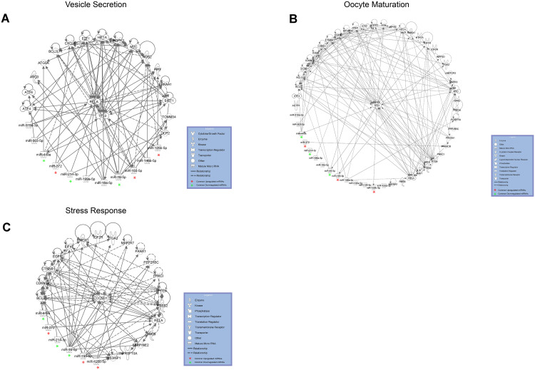
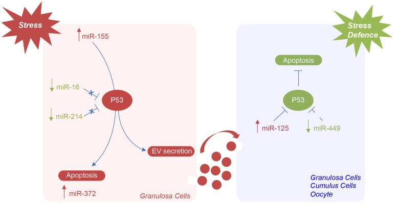
Ovarian aging affects female reproductive potential and is characterized by alterations in proteins, mRNAs and non-coding RNAs inside the ovarian follicle. Ovarian somatic cells and the oocyte communicate with each other secreting different molecules into the follicular fluid, by extracellular vesicles. The cargo of follicular fluid vesicles may influence female reproductive ability; accordingly, analysis of extracellular vesicle content could provide information about the quality of the female germ cell.In order to identify the most significant deregulated microRNAs in reproductive aging, we quantified the small extracellular vesicles in human follicular fluid from older and younger women and analyzed the expression of microRNAs enclosed inside the vesicles. We found twice as many small extracellular vesicles in the follicular fluid from older women and several differentially expressed microRNAs. Correlating microRNA expression profiles with vesicle number, we selected 46 deregulated microRNAs associated with aging. Bioinformatic analyses allowed us to identify six miRNAs involved in TP53 signaling pathways. Specifically, miR-16-5p, miR214-3p and miR-449a were downregulated and miR-125b, miR-155-5p and miR-372 were upregulated, influencing vesicle release, oocyte maturation and stress response. We believe that this approach allowed us to identify a battery of microRNAs strictly related to female reproductive aging.

Keywords: extracellular vesicles; follicular fluid; microRNAs; reproductive aging.


Conflict of interest statement

CONFLICTS OF INTEREST: The authors report no conflicts of interest.

Figures

Figure 1
Figure 1
Morphological and Molecular Characterization of Extracellular Vesicles (EVs) from Follicular Fluid (FF) of older and younger women. (A, B) Scanning Electron Micrographs of EVs isolated from the FF of older (A) and younger women (B) showing the presence of vesicles of spherical shape with a higher abundance in FFs from older women. (C) Diameter distribution of EVs from FFs of older and younger women. Gauss fit of the diameters measured on SEM microscopies shows an EV average diameter of 149 ± 50 nm for FFs from older women (red curve) and of 142 ± 20 nm for those of FFs from younger women (blue trend). (D) The TEM analysis shows that a small Gold (Au) nanoparticle functionalized with an antibody specific for the CD81 protein marker binds to the membrane of small EVs from the FF of younger women.
Figure 2
Figure 2
DLS and NTA analysis. (A) EV size characterization by DLS and NTA. No variation is observed for the EV size in older and younger women. A small difference in the average diameters of EVs is generally found between DLS (where the hydrodynamic radius is measured) and SEM and NTA (that measure the actual radius). (B) Dynamic Light Scattering (DLS) measurements and Nanoparticle tracking analysis (NTA) of small EVs from FFs of older and younger women. Scattered light Intensity, expressed as Kilocounts-per-second (Kcps), and nanoparticle concentration for FFs are shown as red and blue bars respectively. (C) Significant positive linear relationship between DLS and NTA measurements on FF samples. Pearson’s coefficient of correlation (r), R2, and P-value are reported. (D) DLS and (E) NTA show a significantly higher concentration of exosomes in FFs from older women. Kcps and concentration differences related to small EVs of younger and older women are reported as Box plots with Whiskers. The ratio of small EV concentrations ® between younger and older women for DLS and NTA is shown. ** P-values<0.01; * P-value <0.05.
Figure 3
Figure 3
Comparison of DE miRNA sets obtained by two normalization methods for RT-qPCR. Differential expression analysis of miRNAs from FF EVs, comparing older women to younger women and using two normalization methods revealed 64 DE miRNAs (gFC), without considering the number of EVs, and 80 DE miRNAs (vFC) taking into account the number of vesicles. Comparison between gFC and vFC datasets identified 4 classes of DE miRNAs (A-D) for which we show a possible distribution (represented by vertical bars) within the individual EVs from the FF of the two groups of women according to an EV ratio of 2:1 (older vs younger).
Figure 4
Figure 4
GO enrichment analysis for 46 DE common miRNAs identified by two normalization methods. Bar chart representing the most highly statistically enriched Gene Ontologies, in terms of Biological Processes, for DE miRNA targets in female reproductive aging. The x-axis represents the -log10(P-value). The number of target genes in each GO category is shown.
Figure 5
Figure 5
Signaling Pathway enrichment analysis for 46 DE common miRNAs with KEGG. The x-axis represents the -log10(P-value). The number of target genes in each molecular pathway is shown.
Figure 6
Figure 6
DE miRNAs with female aging control target mRNAs within complex regulatory networks of (A) vesicle secretion, (B) oocyte maturation and (C) stress response. DE common miRNAs are highlighted.
Figure 7
Figure 7
Hypothetical model of miRNAs mediating the regulation of TP53 in the ovarian follicle in female reproductive aging. Potential mechanisms by which specific ovarian follicle cell subpopulations can send and receive stress signaling, via EVs, in ovarian aging. MiR-155 expression is induced by stress in granulosa cells. TP53 activation, mediated by the downregulation of miR-16 and miR-214, can initiate apoptosis or lead to cellular senescence and the activation of miR-372 or cause the increase of EV secretion. The upregulation of miR-125 and the downregulation of miR-449 could represent a defense mechanism implemented by follicular cells and oocyte to repress TP53 expression and stress-induced apoptosis.

Similar articles

Cited by

References

    1. López-Otín C, Blasco MA, Partridge L, Serrano M, Kroemer G. The hallmarks of aging. Cell. 2013; 153:1194–217. 10.1016/j.cell.2013.05.039 - DOI - PMC - PubMed
    1. Shirasuna K, Iwata H. Effect of aging on the female reproductive function. Contracept Reprod Med. 2017; 2:23. 10.1186/s40834-017-0050-9 - DOI - PMC - PubMed
    1. Santonocito M, Guglielmino MR, Vento M, Ragusa M, Barbagallo D, Borzì P, Casciano I, Scollo P, Romani M, Tatone C, Purrello M, Di Pietro C. The apoptotic transcriptome of the human MII oocyte: characterization and age-related changes. Apoptosis. 2013; 18:201–11. 10.1007/s10495-012-0783-5 - DOI - PubMed
    1. Webster A, Schuh M. Mechanisms of aneuploidy in human eggs. Trends Cell Biol. 2017; 27:55–68. 10.1016/j.tcb.2016.09.002 - DOI - PubMed
    1. Andronico F, Battaglia R, Ragusa M, Barbagallo D, Purrello M, Di Pietro C. Extracellular vesicles in human oogenesis and implantation. Int J Mol Sci. 2019; 20:2162. 10.3390/ijms20092162 - DOI - PMC - PubMed

Publication types

MeSH terms