Review
. 2020 Aug 6;5(15):e140711.
doi: 10.1172/jci.insight.140711.

The complement system in COVID-19: friend and foe?

Affiliations
Review

The complement system in COVID-19: friend and foe?

Anuja Java et al. JCI Insight. .

Abstract

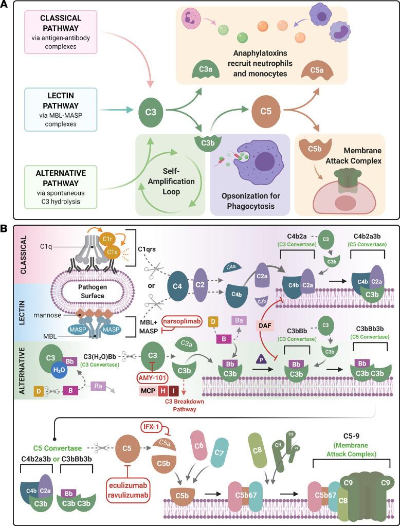
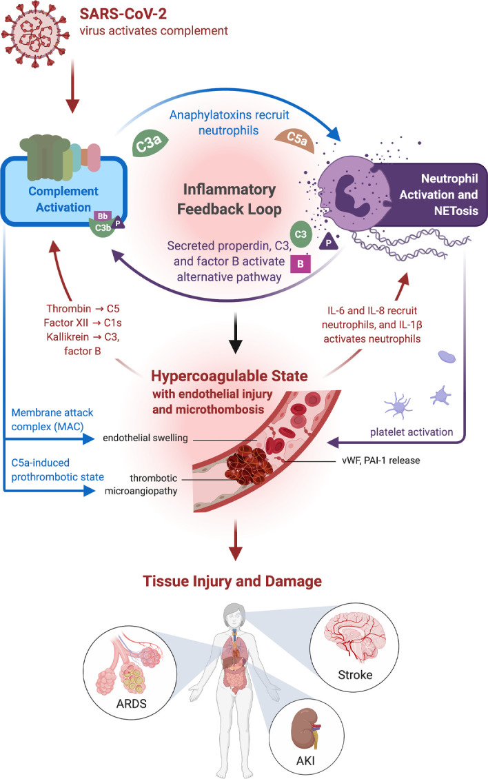
Coronavirus disease 2019 (COVID-19), the disease caused by severe acute respiratory syndrome coronavirus-2 (SARS-CoV-2) has resulted in a global pandemic and a disruptive health crisis. COVID-19-related morbidity and mortality have been attributed to an exaggerated immune response. The role of complement activation and its contribution to illness severity is being increasingly recognized. Here, we summarize current knowledge about the interaction of coronaviruses with the complement system. We posit that (a) coronaviruses activate multiple complement pathways; (b) severe COVID-19 clinical features often resemble complementopathies; (c) the combined effects of complement activation, dysregulated neutrophilia, endothelial injury, and hypercoagulability appear to be intertwined to drive the severe features of COVID-19; (d) a subset of patients with COVID-19 may have a genetic predisposition associated with complement dysregulation; and (e) these observations create a basis for clinical trials of complement inhibitors in life-threatening illness.

Trial registration: ClinicalTrials.gov NCT04369469.


Conflict of interest statement

Conflict of interest: AJ reports serving on the scientific advisory boards of Alexion Pharmaceuticals and Novartis Pharmaceuticals and serving as a consultant for Gemini Therapeutics. JPA reports serving as a consultant for Celldex Therapeutics, Clinical Pharmacy Services, Kypha Inc., Achillion Pharmaceuticals Inc., and BioMarin Pharmaceutical Inc. and stock or equity options in Compliment Corporation, Kypha Inc., Gemini Therapeutics, and AdMiRx Inc. AHJK reports personal fees (<$10,000) from Exagen Diagnostics Inc. and GlaxoSmithKline.

Figures

Figure 1
Figure 1. Schematic diagram of the activation of the 3 complement cascades and COVID-19 therapeutically targeted components.
A simplified (A) and detailed (B) description of the complement cascade are shown. There are 3 major independent, but overlapping, pathways for activation of complement. The classical, lectin, and alternative pathways generate the major complement opsonin C3b and the membrane attack complex (MAC, C5b-9). The engagement of the C1 complex (C1qrs) with antigen-antibody complexes or pentraxins generates bimolecular C3 convertase (C4b2a). The latter cleaves C3 to C3b and releases the anaphylatoxin, C3a. The binding of another C3b to the convertase generates the C5 convertase (C4b2a3b). The lectin pathway is similar, except mannose-associated serine proteases (MASPs) substitute for C1 proteases (C1r and C1s). The AP self-activates by the low-level turnover of C3 in serum to C3(H2O). This nondiscriminate tickover, particularly on a pathogen surface or damaged tissue, rapidly engages factors B (B) and D (D) to form a C3 convertase C3(H2O)Bb to trigger more C3b. C3b generated by any one of the 3 pathways opsonizes the target, binds a C3 convertase to generate the trimolecular C5 convertases (C4b2a3b or C3bBbC3b), and amplifies more C3b via the AP. Cleavage of C5 releases the potent anaphylatoxin C5a, while the C5b initiates the terminal pathway to form the MAC. C3a and C5a are potent chemoattractants for neutrophils and monocytes. Membrane-bound (CD46, CD55) and fluid-phase regulators (factor H [FH], factor I [FI]) prevent fluid-phase activation and activation on normal cells and tissues. For example, membrane cofactor protein (MCP, also known as CD46) serves as a cofactor the FI-mediated inactivation of C3b to iC3b and C4b to C4c and C4d. The current armamentarium of complement inhibitors being tested in COVID-19 targets different aspects of this cascade. AMY-101 is a synthetic peptide that inhibits C3, whereas both eculizumab and ravulizumab are humanized monoclonal antibodies against C5. IFX-1 is a monoclonal antibody specifically targeting C5a, and narsoplimab is a human monoclonal antibody targeting MASP-2.
Figure 2
Figure 2. A summary of SARS-CoV-2 and complement activation leading to immune hyperinflammatory reactions and resulting in human pathology.
Complement activation generates the proinflammatory polypeptides, C3a and C5a, and recruits neutrophils as well as monocytes. Activated neutrophils generate web-like extracellular traps (NETs), in a process known as NETosis, that contain components such as C3, properdin (P), and factor B (B) that activate the alternative complement pathway and engage an inflammatory feedback loop. Although NETs assist in host defense against pathogens, a sustained response, such as that seen in COVID-19, may incite ongoing inflammation and a hypercoagulable state. Additionally, the membrane attack complex (MAC) also induces endothelial inflammation and tissue injury, leading to the generation of IL-6 and IL-1β, which continue to propagate NETosis. Endothelial injury leads to the generation of vWF multimers. Excess ultralong vWF stabilizes factor VIII activity and prevents the binding of factor I. Endothelial damage also results in the release of plasminogen activator inhibitor-1 (PAI-1), which exacerbates thrombosis, along with C5a-induced release of tissue factor and other prothrombotic proteins. These changes then can augment a complement-coagulation pathway crosstalk, due to serine proteases, such as thrombin and kallikrein, activating the complement system in a convertase-independent manner. Such interactions among endothelial injury, hypercoagulability, and complement activation cause tissue damage, such as acute respiratory distress syndrome (ARDS), acute kidney injury (AKI), and stroke, and are often associated with a thrombotic microangiopathy.

Similar articles

  • Immune complement and coagulation dysfunction in adverse outcomes of SARS-CoV-2 infection.
    Ramlall V, Thangaraj PM, Meydan C, Foox J, Butler D, Kim J, May B, De Freitas JK, Glicksberg BS, Mason CE, Tatonetti NP, Shapira SD. Ramlall V, et al. Nat Med. 2020 Oct;26(10):1609-1615. doi: 10.1038/s41591-020-1021-2. Epub 2020 Aug 3. Nat Med. 2020. PMID: 32747830 Free PMC article.
  • Severe COVID-19: what have we learned with the immunopathogenesis?
    Bordallo B, Bellas M, Cortez AF, Vieira M, Pinheiro M. Bordallo B, et al. Adv Rheumatol. 2020 Sep 22;60(1):50. doi: 10.1186/s42358-020-00151-7. Adv Rheumatol. 2020. PMID: 32962761 Free PMC article.
  • Neutrophils and Contact Activation of Coagulation as Potential Drivers of COVID-19.
    Busch MH, Timmermans SAMEG, Nagy M, Visser M, Huckriede J, Aendekerk JP, de Vries F, Potjewijd J, Jallah B, Ysermans R, Oude Lashof AML, Breedveld PH, van de Poll MCG, van de Horst ICC, van Bussel BCT, Theunissen ROMFIH, Spronk HMH, Damoiseaux JGMC, Ten Cate H, Nicolaes GAF, Reutelingsperger CP, van Paassen P. Busch MH, et al. Circulation. 2020 Nov 3;142(18):1787-1790. doi: 10.1161/CIRCULATIONAHA.120.050656. Epub 2020 Sep 18. Circulation. 2020. PMID: 32946302 Free PMC article. No abstract available.
  • Systemic complement activation is associated with respiratory failure in COVID-19 hospitalized patients.
    Holter JC, Pischke SE, de Boer E, Lind A, Jenum S, Holten AR, Tonby K, Barratt-Due A, Sokolova M, Schjalm C, Chaban V, Kolderup A, Tran T, Tollefsrud Gjølberg T, Skeie LG, Hesstvedt L, Ormåsen V, Fevang B, Austad C, Müller KE, Fladeby C, Holberg-Petersen M, Halvorsen B, Müller F, Aukrust P, Dudman S, Ueland T, Andersen JT, Lund-Johansen F, Heggelund L, Dyrhol-Riise AM, Mollnes TE. Holter JC, et al. Proc Natl Acad Sci U S A. 2020 Oct 6;117(40):25018-25025. doi: 10.1073/pnas.2010540117. Epub 2020 Sep 17. Proc Natl Acad Sci U S A. 2020. PMID: 32943538 Free PMC article.
  • COVID-19: Complement, Coagulation, and Collateral Damage.
    Lo MW, Kemper C, Woodruff TM. Lo MW, et al. J Immunol. 2020 Sep 15;205(6):1488-1495. doi: 10.4049/jimmunol.2000644. Epub 2020 Jul 22. J Immunol. 2020. PMID: 32699160 Free PMC article. Review.

Cited by

References

    1. Vabret N, et al. Immunology of COVID-19: Current State of the Science. Immunity. 2020;52(6):910–941. doi: 10.1016/j.immuni.2020.05.002. - DOI - PMC - PubMed
    1. Bloch EM, et al. Deployment of convalescent plasma for the prevention and treatment of COVID-19. J Clin Invest. 2020;130(6):2757–2765. doi: 10.1172/JCI138745. - DOI - PMC - PubMed
    1. Angus DC, et al. The REMAP-CAP (Randomized Embedded Multifactorial Adaptive Platform for Community-acquired Pneumonia) Study. Rationale and Design. Ann Am Thorac Soc. 2020;17(7):879–891. doi: 10.1513/AnnalsATS.202003-192SD. - DOI - PMC - PubMed
    1. Coronaviridae Study Group of the International Committee on Taxonomy of Viruses. The species Severe acute respiratory syndrome-related coronavirus: classifying 2019-nCoV and naming it SARS-CoV-2. Nat Microbiol. 2020;5(4):536–544. doi: 10.1038/s41564-020-0695-z. - DOI - PMC - PubMed
    1. Henrickson SE. Learning from our immunological history: What can SARS-CoV teach us about SARS-CoV-2? Sci Immunol. 2020;5(46):eabb8618. doi: 10.1126/sciimmunol.abb8618. - DOI - PubMed

Publication types

Associated data