. 2020 Aug 19;82(8):1113-1117.
doi: 10.1292/jvms.20-0159. Epub 2020 Jun 17.

Decreased Anti-Müllerian hormone and Anti-Müllerian hormone receptor type 2 in hypothalami of old Japanese Black cows

Affiliations

Decreased Anti-Müllerian hormone and Anti-Müllerian hormone receptor type 2 in hypothalami of old Japanese Black cows

Onalenna Kereilwe et al. J Vet Med Sci. .

Abstract

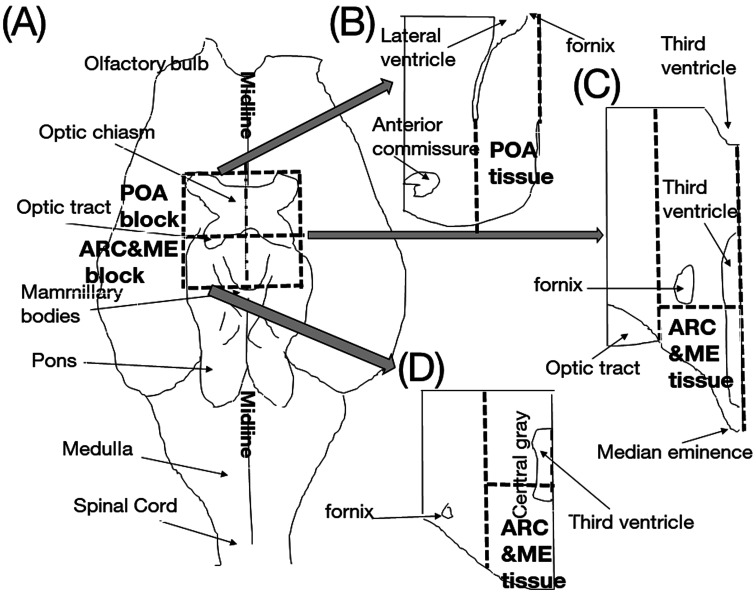
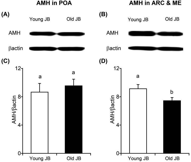
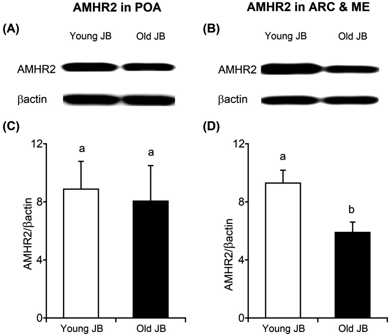
Cow fertility decreases with age, but the hypothalamic pathomechanisms are not understood. Anti-Müllerian hormone (AMH) stimulates gonadotropin-releasing hormone (GnRH) neurons via AMH receptor type 2 (AMHR2), and most GnRH neurons in the preoptic area (POA), arcuate nucleus (ARC), and median eminence (ME) express AMH and AMHR2. Therefore, we hypothesized that both protein amounts would differ in the anterior hypothalamus (containing the POA) and posterior hypothalamus (containing the ARC and ME) between young post-pubertal heifers and old cows. Western blot analysis showed lower (P<0.05) expressions of AMH and AMHR2 in the posterior hypothalamus, but not in the anterior hypothalamus, of old Japanese Black cows compared to young heifers. Therefore, AMH and AMHR2 were decreased in the posterior hypothalami of old cows.

Keywords: Müllerian inhibiting substance; female reproductive senescence; gonadotropin-releasing hormone neuron; preoptic area; ruminant.


Figures

Fig. 1.
Fig. 1.
Schematic illustration of brain tissue sampling according to a previous study [6]. Briefly, brain tissues were dissected as per the dotted line on the ventral side (A) with the following margins: rostrally-rostral border of the optic chiasm; caudally-rostral to the mammillary bodies; lateral to the optic chiasm; and 0.5 cm dorsal to the third ventricle. We then split the block into two parts by cutting rostral to the median eminence (ME), yielding an anterior part containing the preoptic area (POA block) and a posterior part containing the arcuate nucleus and median eminence (ARC&ME block) according to a method reported by Hassaneen et al [3]. The blocks were further cut using a previously reported method [6]. Both the POA and ARC&ME blocks were cut at the midlines to obtain left and right sides. Using the bovine brain atlas [10, 14] as a reference, the blocks were further cut using their exterior shapes and the third or lateral ventricles as landmarks as the dotted line (B, C, D). Finally, the size of each tissue sample containing the preoptic area (POA tissue) was less than 1 cm along its lateral axis; 2 cm along the rostrocaudal axis; and 3 cm along the vertical axis. The size of each tissue containing the arcuate nucleus and median eminence (ARC&ME tissue) was less than 1 cm along its lateral axis; 2 cm along the rostrocaudal axis; and 1 cm along the vertical axis.
Fig. 2.
Fig. 2.
Representative Anti-Müllerian hormone (AMH) (in mature C-terminal form) and β-actin immunoreactive protein bands in preoptic area (POA; A), and arcuate nucleus and median eminence (ARC&ME; B) tissues obtained from Japanese black young heifers and old cows. Comparison of AMH protein expression normalized to that of β-actin in young heifers (n=5) and old cows (n=5) in POA (C) and ARC&ME (D). Letters (a vs. b) indicate significant differences (P<0.05) between groups.
Fig. 3.
Fig. 3.
Representative AMH receptor type 2 (AMHR2) and β-actin immunoreactive protein bands in preoptic area (POA; A), and arcuate nucleus and median eminence (ARC&ME; B) tissues obtained from young heifers and old cows. Comparison of AMHR2 protein expression level normalized to β-actin in young heifers (n=5) and old cows (n=5) in POA (C) and ARC&ME (D). Letters (a vs. b) indicate significant differences (P<0.05) between groups.

Similar articles

Cited by

References

    1. Bond B. C., Virley D. J., Cairns N. J., Hunter A. J., Moore G. B., Moss S. J., Mudge A. W., Walsh F. S., Jazin E., Preece P.2002. The quantification of gene expression in an animal model of brain ischaemia using TaqMan real-time RT-PCR. Brain Res. Mol. Brain Res. 106: 101–116. doi: 10.1016/S0169-328X(02)00417-5 - DOI - PubMed
    1. Cimino I., Casoni F., Liu X., Messina A., Parkash J., Jamin S. P., Catteau-Jonard S., Collier F., Baroncini M., Dewailly D., Pigny P., Prescott M., Campbell R., Herbison A. E., Prevot V., Giacobini P.2016. Novel role for anti-Müllerian hormone in the regulation of GnRH neuron excitability and hormone secretion. Nat. Commun. 7: 10055. doi: 10.1038/ncomms10055 - DOI - PMC - PubMed
    1. Hassaneen A., Naniwa Y., Suetomi Y., Matsuyama S., Kimura K., Ieda N., Inoue N., Uenoyama Y., Tsukamura H., Maeda K. I., Matsuda F., Ohkura S.2016. Immunohistochemical characterization of the arcuate kisspeptin/neurokinin B/dynorphin (KNDy) and preoptic kisspeptin neuronal populations in the hypothalamus during the estrous cycle in heifers. J. Reprod. Dev. 62: 471–477. doi: 10.1262/jrd.2016-075 - DOI - PMC - PubMed
    1. Jansen H. T., Hileman S. M., Lubbers L. S., Kuehl D. E., Jackson G. L., Lehman M. N.1997. Identification and distribution of neuroendocrine gonadotropin-releasing hormone neurons in the ewe. Biol. Reprod. 56: 655–662. doi: 10.1095/biolreprod56.3.655 - DOI - PubMed
    1. Kamomae H.2012. Reproductive disturbance. pp. 283–340. In: Veterinary Theriogenology (Nakao, T., Tsumagari, S. and Katagiri, S. eds.), Buneidou Press, Tokyo (in Japanese).

MeSH terms

Substances