

The Shape of Posterior Sclera as a Biometric Signature in Open-angle Glaucoma: An Intereye Comparison Study
- PMID: 32555059
- PMCID: PMC7647446
- DOI: 10.1097/IJG.0000000000001573
The Shape of Posterior Sclera as a Biometric Signature in Open-angle Glaucoma: An Intereye Comparison Study
Abstract
Purpose: To characterize intereye differences in posterior segment parameters and determine their significance in open-angle glaucoma patients with unilateral damage.
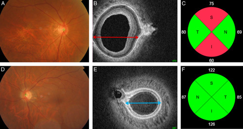
Methods: Both eyes from 65 subjects without any nerve damage and 43 patients undergoing treatment for unilateral open-angle glaucoma were included in this study. A 12.0×9.0×2.6 mm volume of the posterior segment in each eye was scanned with swept-source optical coherence tomography. Coronally reconstructed optical coherence tomography images were analyzed to determine the deepest point of the eye (DPE), which we then calculated the distance (Disc-DPE distance), depth (Disc-DPE depth), angle (Disc-DPE angle) from the optic disc center to the DPE. Posterior pole shape was analyzed measuring the posterior pole-cross-sectional area, posterior pole-horizontal width (PP-HW), and posterior pole-vertical width) of the posterior pole. These measurements and their intereye absolute difference (IAD; absolute difference in measurements between the right and left eyes) values were compared between the healthy and unilateral glaucomatous patients.
Results: The posterior sclera measurements, including the Disc-DPE distance, Disc-DPE depth, and posterior pole-cross-sectional area, were significantly different between the unilateral glaucoma eyes and contralateral healthy eyes (P=0.043, P=0.035, and P=0.049, respectively). By contrast, none of the intereye differences in optic nerve head parameters were significant in the unilateral glaucoma patients. In comparison with the IAD values, the baseline intraocular pressure and PP-HW of the posterior segment showed significant differences between the healthy and the unilateral glaucoma patients (P=0.019 and P=0.036, respectively). A multivariate analysis showed that a larger baseline intraocular pressure IAD [odds ratio (OR), 1.381; P=0.009)] and larger PP-HW IAD (OR, 1.324; P=0.032) were significantly associated with the presence of glaucoma.
Conclusions: Compared with the fellow healthy eyes, glaucomatous eyes had larger and more steeply curved posterior poles, which represent a structural variation of the posterior sclera that might be associated with glaucomatous optic neuropathy.
Conflict of interest statement
Disclosure: The authors declare no conflict of interest.
Figures


References
-
- Boland MV, Quigley HA. Risk factors and open-angle glaucoma: classification and application. J Glaucoma. 2007;16:406–418. - PubMed
-
- Anderson DR, Drance SM, Schulzer M. Collaborative Normal-Tension Glaucoma Study G. Natural history of normal-tension glaucoma. Ophthalmology. 2001;108:247–253. - PubMed
-
- Heijl A, Bengtsson B, Hyman L, et al. Early Manifest Glaucoma Trial G. Natural history of open-angle glaucoma. Ophthalmology. 2009;116:2271–2276. - PubMed
-
- Burgoyne CF, Downs JC, Bellezza AJ, et al. The optic nerve head as a biomechanical structure: a new paradigm for understanding the role of IOP-related stress and strain in the pathophysiology of glaucomatous optic nerve head damage. Prog Retin Eye Res. 2005;24:39–73. - PubMed
Publication types
MeSH terms
LinkOut - more resources
Full Text Sources
Miscellaneous
