. 2020 Jun 18;11(6):473.
doi: 10.1038/s41419-020-2674-y.

NORAD orchestrates endometrial cancer progression by sequestering FUBP1 nuclear localization to promote cell apoptosis

Affiliations

NORAD orchestrates endometrial cancer progression by sequestering FUBP1 nuclear localization to promote cell apoptosis

Tong Han et al. Cell Death Dis. .

Abstract

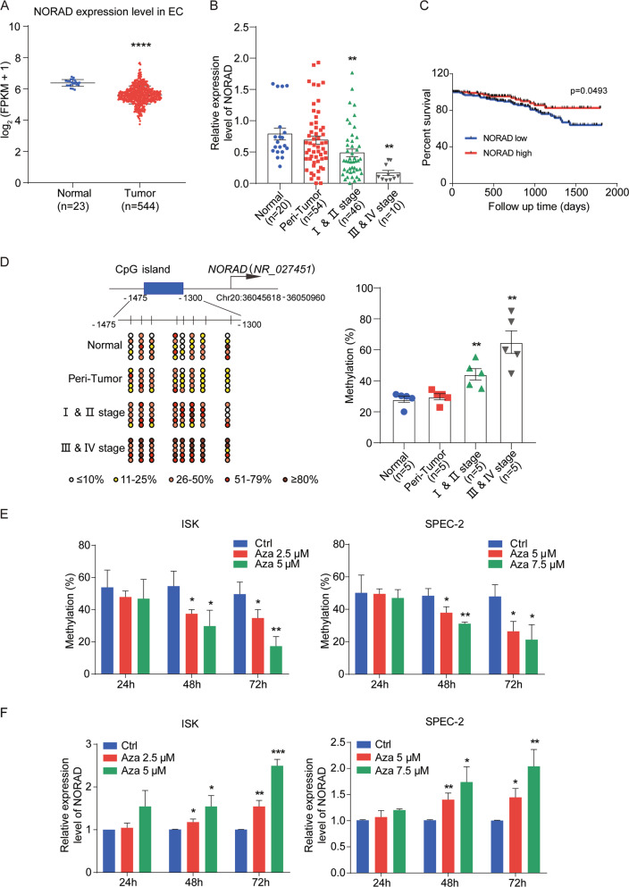
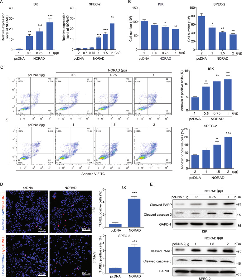
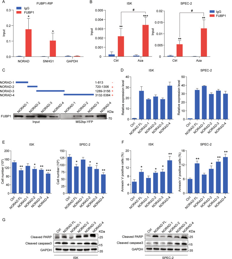
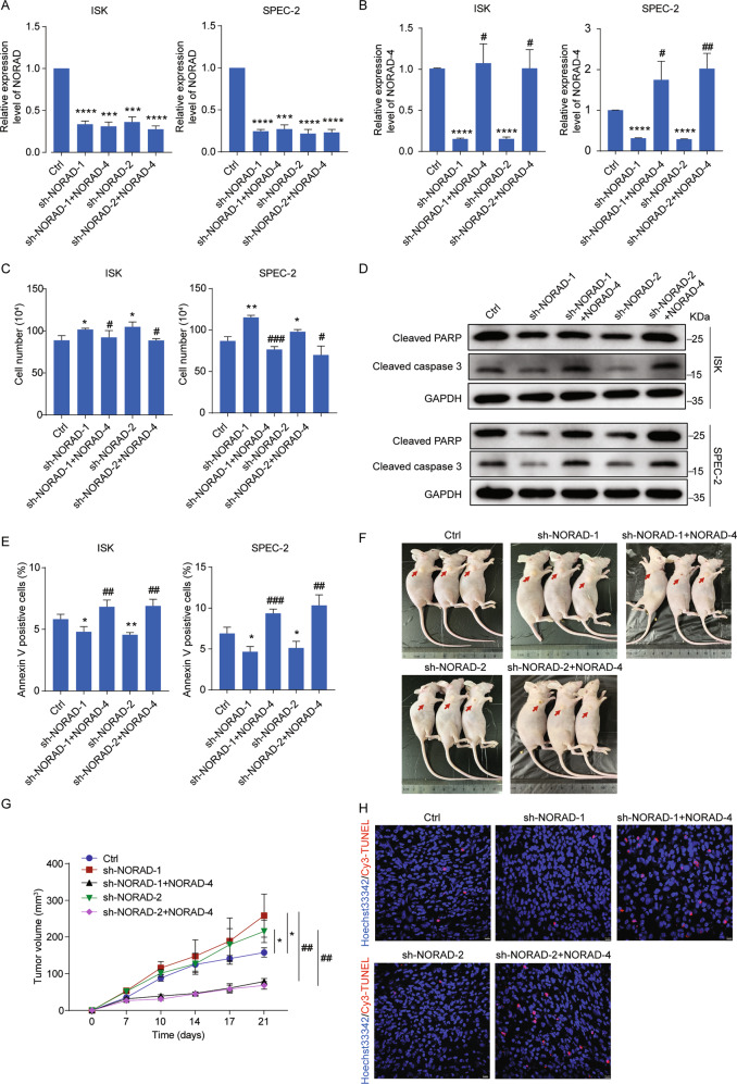
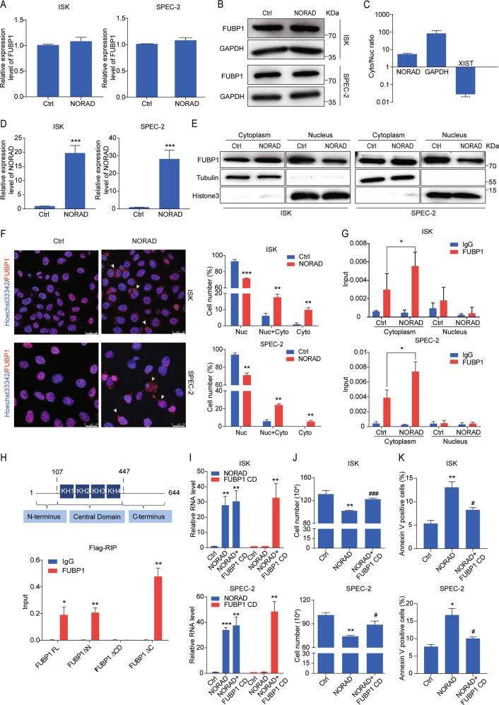
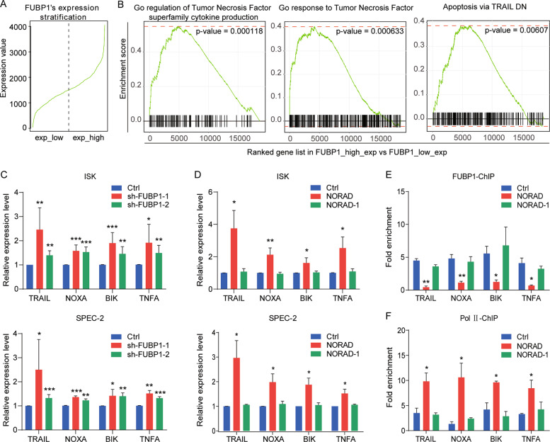
Long noncoding RNAs (lncRNAs) are emerging as critical regulators in tumor initiation and progression. However, the biological mechanisms and potential clinical application of lncRNA NORAD in endometrial cancer (EC) remain unknown. Herein, we identified NORAD underwent promoter hypermethylation-associated downregulation in EC. Epigenetic inactivation of NORAD was correlated with EC progression (FIGO stage) and poor outcome. Overexpression of NORAD significantly inhibited cell growth and promoted apoptosis in EC cells. Mechanistic studies revealed that multiple regions of NORAD served as a platform for binding with the central domain of anti-apoptotic factor FUBP1. Our findings further indicated that the NORAD/FUBP1 interaction attenuated FUBP1 nuclear localization and thus impaired the occupancies of FUBP1 on its target pro-apoptotic gene promoters, resulting in apoptosis induction in EC. Moreover, knockdown of NORAD promoted tumor growth in the xenograft mice model. While, introduction of NORAD-4 fragment, which bound with FUBP1, successfully reversed tumor growth and apoptosis inhibition mediated by NORAD knockdown in vivo. Our findings provide mechanistic insight into the critical roles of NORAD as a tumor suppressor in EC progression. NORAD could possibly serve as a novel prognostic biomarker and provide the rationale for EC therapy.


Conflict of interest statement

The authors declare that they have no conflict of interest.

Figures

Fig. 1
Fig. 1. Downregulation of NORAD due to promoter hypermethylation is correlated with progression and prognosis of EC.
a Relative NORAD expression in the EC patient cohort compared with that in normal endometrial tissues according to TCGA dataset. b The NORAD expression level in 20 normal endometrial tissues, 56 EC (including 46 I & II stage and 10 III & IV stage patients), and 54 peri-tumor tissues was detected by qRT-PCR. c The survival data from TCGA EC patient cohort containing 308 endometrioid endometrial adenocarcinomas (the major subtype of EC) were analyzed by Kaplan–Meier analysis. d Methylation status of the CpG sites at the promoter of NORAD in normal (n = 5), peri-tumor (n = 5), I & II stage (n = 5), and III & IV stage EC patients (n = 5) was investigated by bisulfite sequencing. The average percentages of unmethylated and methylated CpGs of 10 clones from each patient were presented by different colors according to the methylated degree. e The methylation analysis of NORAD promoter in ISK and SPEC-2 cells with the distinct doses and times of Azacitidine (Aza) treatment, performed by bisulfite sequencing. f Restored expression of NORAD after treatment with Aza in EC cells at different doses and times. The results were determined from triplicates, and the error bars represented as the mean ± SEM in patients’ samples, and the mean ± SD in EC cell lines, *P < 0.05, **P < 0.01, ***P < 0.001, ****P < 0.0001. NORAD noncoding RNA activated by DNA damage, EC endometrial cancer, Aza azacitidine.
Fig. 2
Fig. 2. NORAD promotes apoptosis in EC cells.
a qRT-PCR analysis for the expression level of NORAD in ISK and SPEC-2 EC cell lines with different doses transfection of NORAD, in comparison with the empty vectors. b Cell-counting assays for the control and ectopic NORAD-expressing ISK and SPEC-2 cells after 48 h transfection. c Increased percentage of apoptosis in the ectopic NORAD-expressing EC cells after 48 h transfection via FACS analysis. d TUNEL assays for apoptotic cells in the control and NORAD-expressing EC groups (left). Statistics of the TUNEL-Cy3 positive cells are shown (right). Scale bar, 100 μm. e Activated expression of cleaved PARP and cleaved caspase-3 was visualized by western blot. The results were determined from triplicates, and the error bars represented as the mean ± SD, *P < 0.05, **P < 0.01, ***P < 0.001. TUNEL TdT-mediated dUTP Nick-end labeling, GAPDH glyceraldehyde-3-phosphate dehydrogenase.
Fig. 3
Fig. 3. The binding of NORAD and FUBP1 is essential for NORAD to induce apoptosis.
a qRT-PCR analysis for the binding of NORAD, SNHG1, and GAPDH expression by an anti-FUBP1 antibody compared with that by an IgG control antibody in 293FT cells. SNHG1 and GAPDH served as the positive and negative controls, respectively. b qRT-PCR analysis for the binding of NORAD on FUBP1 in EC cells with Aza treatment by RIP assays. c Western blot for the FUBP1 protein (lower panel) pulled down by truncated NORAD (NORAD-1, -2, -3, and -4; upper panel). d The expression levels of full-length NORAD and fragments transfected in ISK and SPEC-2 cells were detected by qRT-PCR. e Cell-counting assays for ISK and SPEC-2 cells transfected with full-length NORAD and fragments, respectively. f The percentage of apoptotic cells transfected with full-length NORAD and fragments, evaluated by FACS analysis. g Western blot for the expression of cleaved PARP and cleaved caspase-3 after ectopic expression of full-length NORAD and fragments. The results were determined from triplicates, and the error bars represented as the mean ± SD, */# P < 0.05, **P < 0.01, ***P < 0.001. FUBP1 far upstream element-binding protein 1, SNHG1 small nucleolar RNA host gene 1, RIP RNA immunoprecipitation, YFP yellow fluorescent protein.
Fig. 4
Fig. 4. NORAD-4 rescues the apoptosis inhibition and tumor growth mediated by knockdown of NORAD in vitro and in vivo.
a, b The expression level of NORAD (a) and NORAD-4 (b) was detected by qRT-PCR in NORAD knockdown cell lines and in NORAD knockdown rescued by NORAD-4 fragment cell lines. c Cell-counting assays for the NORAD knockdown and rescued by NORAD-4 cell lines. d The expression levels of apoptotic associated markers were detected by western blot in the NORAD knockdown and rescued by NORAD-4 cell lines. e The percentage of apoptotic cells in the NORAD knockdown and rescued by NORAD-4 cell lines was analyzed by FACS. f SPEC-2-derived cell lines (sh-NORAD-1/2, sh-NORAD-1/2 + NORAD-4) were subcutaneously injected into the hind flanks of nude mice. g Tumor volume was monitored from day 0 to day 21 post injection. h Apoptosis in tumor tissues was presented by TUNEL assay. Scale bar, 10 μm. The results were determined from triplicates, and the error bars represented as the mean ± SD, */#P < 0.05, */##P < 0.01, ***/###P < 0.001, ****P < 0.0001.
Fig. 5
Fig. 5. NORAD impairs the nuclear localization of FUBP1 through its central domain.
a, b The FUBP1 expression level after the introduction of NORAD in ISK and SPEC-2 cells via qRT-PCR (a) and western blot (b). c The subcellular distribution of NORAD was analyzed by qRT-PCR. GAPDH and XIST genes were used as controls for the cytoplasmic and nuclear fractions, respectively. d The expression level of NORAD was detected by qRT-PCR. e The fractionation of FUBP1 was visualized by western blot after ectopic expression of NORAD in ISK and SPEC-2 cells. GAPDH and Histone 3 indicated the cytoplasmic and nuclear fractions, respectively. f Immunofluorescence assays indicated the altered localization of FUBP1 (red) after introduction of NORAD in ISK and SPEC-2 cells (left). Quantifications of the percentages of FUBP1 presented only in the nucleus, in the cytoplasm, and both in the nucleus and cytoplasm are shown (right). White arrows represented the cells which FUBP1 was distributed both in the cytoplasm and nucleus. Yellow arrows represented the cells in which FUBP1 was distributed only in the cytoplasm. Scale bar, 25 μm. g The subcellular fractionation followed by RIP assays was performed to analyze the interaction of NORAD and FUBP1 in the cytoplasmic and nuclear lysates of NORAD overexpressing cells. h qRT-PCR analysis of NORAD immunoprecipitated by Flag-tagged full-length and three deleted mutations of FUBP1 in 293FT cells compared with the IgG control. i The expression level of NORAD and FUBP1 CD truncation was detected by qRT-PCR. j Cell-counting assays of the rescued cell growth by FUBP1 CD in the NORAD-expressing ISK and SPEC-2 cells. k FACS analysis of the rescued percentage of apoptotic cells by FUBP1 CD in the NORAD-expressing ISK and SPEC-2 cells. The results were determined from triplicates, and the error bars represented as the mean ± SD, */#P < 0.05, **P < 0.01, ***/###P < 0.001. XIST X inactivation-specific transcript.
Fig. 6
Fig. 6. The NORAD/FUBP1 interaction results in the upregulation of downstream pro-apoptotic genes.
a RNA-seq data of liver cancer from TCGA to classify FUBP1 high- and low-expressed groups. b GSEA for the FUBP1-related pathways in liver cancer. c qRT-PCR analysis for the expression of four FUBP1 downstream targets (TRAIL, NOXA, BIK, and TNFA) by knockdown of FUBP1. d qRT-PCR analysis for the expression of four FUBP1 downstream target genes by the introduction of full-length NORAD or the NORAD-1 fragment, which was not bound to FUBP1. e, f ChIP-qPCR analysis for the FUBP1 (e) and RNA polymerase II (f) occupancies at the promoters of four target genes (TRAIL, NOXA, BIK, and TNFA) after transfecting full-length NORAD or NORAD-1 fragment. The fold enrichment was relative to the input DNA. The results were determined from triplicates, and the error bars represented as the mean ± SD, *P < 0.05, **P < 0.01, ***P < 0.001. TNF tumor necrosis factor, TRAIL TNF-related apoptosis-inducing ligand, NOXA PMAIP1, phorbol-12-myristate-13-acetate-induced protein 1, BIK BCL2 interacting killer, ChIP chromatin immunoprecipitation, Pol II RNA polymerase II.

Similar articles

Cited by

References

    1. Siegel RL, Miller KD, Jemal A. Cancer statistics, 2020. CA Cancer J. Clin. 2020;70:7–30. - PubMed
    1. Brooks RA, et al. Current recommendations and recent progress in endometrial cancer. CA Cancer J. Clin. 2019;69:258–279. - PubMed
    1. Morice P, Leary A, Creutzberg C, Abu-Rustum N, Darai E. Endometrial cancer. Lancet. 2016;387:1094–1108. - PubMed
    1. Schmitt AM, Chang HY. Long noncoding RNAs in cancer pathways. Cancer Cell. 2016;29:452–463. - PMC - PubMed
    1. Gutschner T, Diederichs S. The hallmarks of cancer: a long non-coding RNA point of view. RNA Biol. 2012;9:703–719. - PMC - PubMed

Publication types

MeSH terms