. 2020 Jun 18;11(6):472.
doi: 10.1038/s41419-020-2581-2.

Long noncoding RNA DLEU2 predicts a poor prognosis and enhances malignant properties in laryngeal squamous cell carcinoma through the miR-30c-5p/PIK3CD/Akt axis

Affiliations

Long noncoding RNA DLEU2 predicts a poor prognosis and enhances malignant properties in laryngeal squamous cell carcinoma through the miR-30c-5p/PIK3CD/Akt axis

Xiaoming Li et al. Cell Death Dis. .

Abstract

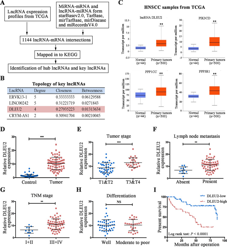
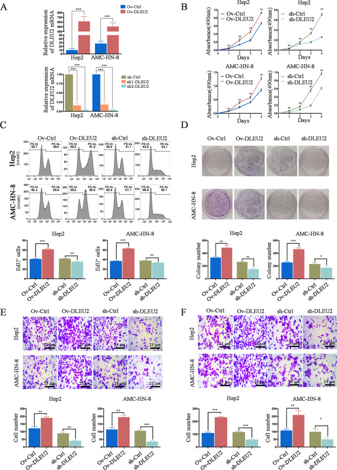
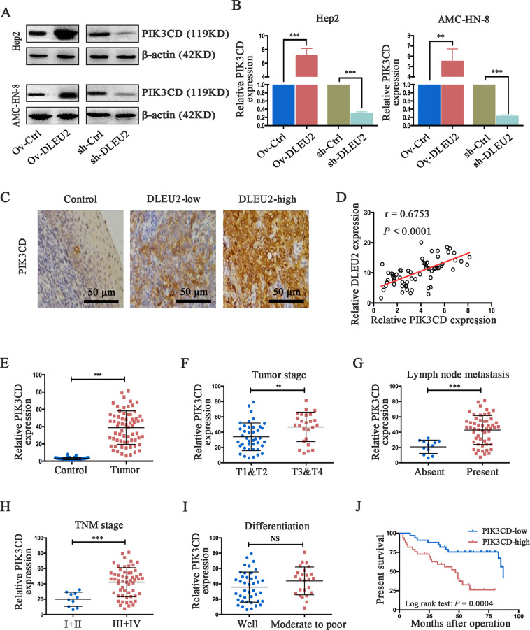
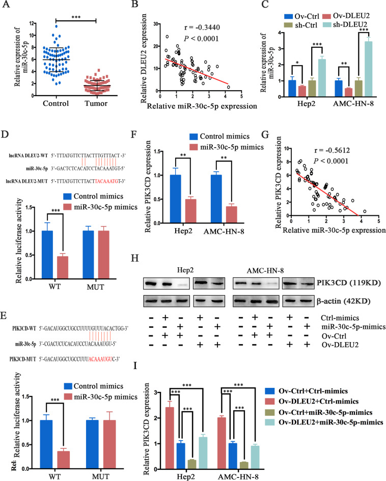
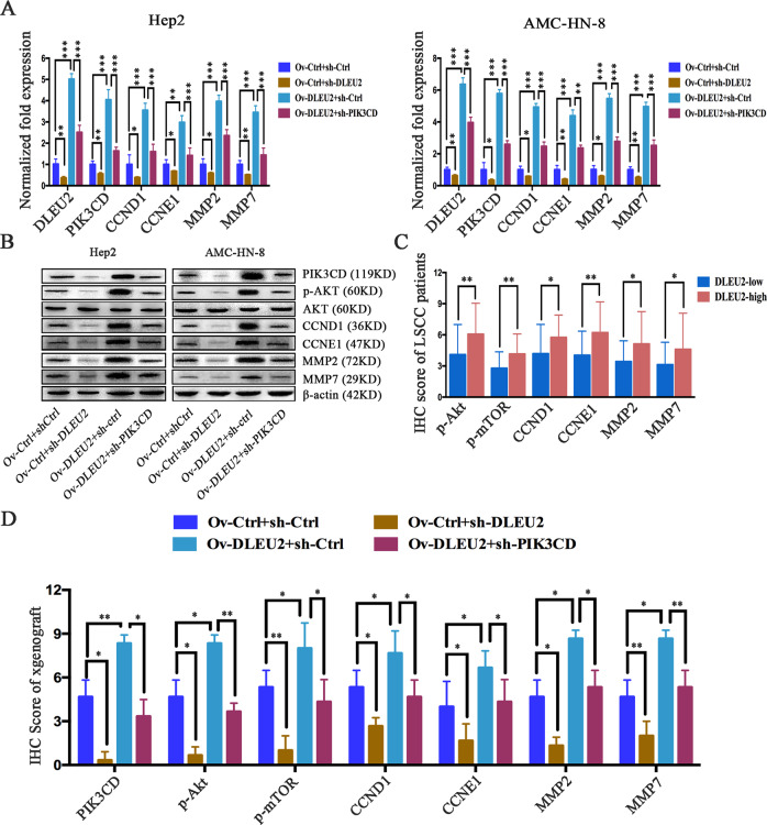
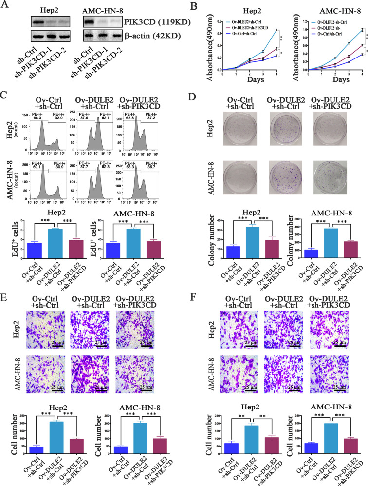
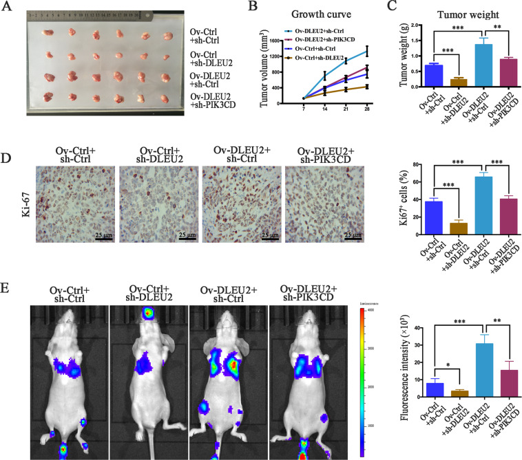
Long noncoding RNAs (lncRNAs) have been identified as potential prognostic tools and therapeutic biomarkers for a variety of human cancers. However, the functional roles and underlying mechanisms of key lncRNAs affecting laryngeal squamous cell carcinomas (LSCCs) are largely unknown. Here, we adopted a novel subpathway strategy based on the lncRNA-mRNA profiles from the Cancer Genome Atlas (TCGA) database and identified the lncRNA deleted in lymphocytic leukemia 2 (DLEU2) as an oncogene in the pathogenesis of LSCCs. We found that DLEU2 was significantly upregulated and predicted poor clinical outcomes in LSCC patients. In addition, ectopic overexpression of DLEU2 promoted the proliferation and migration of LSCC cells both in vivo and in vitro. Mechanistically, DLEU2 served as a competing endogenous RNA to regulate PIK3CD expression by sponging miR-30c-5p and subsequently activated the Akt signaling pathway. As a target gene of DLEU2, PIK3CD was also upregulated and could predict a poor prognosis in LSCC patients. In conclusion, we found that the novel LSCC-related gene DLEU2 enhances the malignant properties of LSCCs via the miR-30c-5p/PIK3CD/Akt axis. DLEU2 and its targeted miR-30c-5p/PIK3CD/Akt axis may represent valuable prognostic biomarkers and therapeutic targets for LSCCs.


Conflict of interest statement

The authors declare that they have no conflict of interest.

Figures

Fig. 1
Fig. 1. DLEU2 is a novel LSCC-related oncogene and predicts the prognosis in LSCC patients.
a Schematic depicting key lncRNAs and hub genes in the LRSP subpathways and network. b Topology features of the key lncRNAs. c DLEU2 and its potential target genes (PIK3CD, PPP1CC, and PPP3R1) were significantly upregulated in the tumor tissues compared with the normal control tissues in HNSCC patients from the TCGA database. d The mRNA expression of DLEU2 was evaluated by qRT-PCR analysis in 66 cases of LSCC specimens and the matched adjacent normal tissues. e–h Correlations of DLEU2 expression with the clinicopathological characteristics of the tumor stage (e), lymph node metastasis (f), tumor node metastasis (g), and differentiation (h) in LSCC specimens. i The correlation between survival time and DLEU2 expression was analyzed in LSCC patients. **P < 0.01, ***P < 0.001.
Fig. 2
Fig. 2. DLEU2 modulates LSCC cell proliferation, invasion, and migration in vitro.
a The efficiency of transfection was detected by qRT-PCR, as indicated. b The CCK-8 assay was performed to evaluate the cell viability at various time points in the indicated cells. c, d The EdU proliferation assay (c) and colony formation assay (d) were performed to evaluate the effect of DLEU2 on the proliferation of the indicated cells. e, f The transwell assay was performed to evaluate the invasion (e) and migration (f) abilities of the indicated cells (original magnification: ×200; scale bar = 25 µm). The results are presented as the mean ± SD from three independent experiments. *P < 0.05, **P < 0.01, ***P < 0.001.
Fig. 3
Fig. 3. DLEU2 regulates PIK3CD expression, which is correlated with tumor progression and survival time in LSCC patients.
a, b DLEU2 was overexpressed or knocked down in LSCC cells. Western blot (a) and qRT-PCR (b) analysis were performed to detect PIK3CD expression. β-actin was used as an internal control. c Representative immunohistochemical staining of PIK3CD in normal control, low DLEU2 expression, and high DLEU2 expression samples, respectively (original magnification: ×200; scale bar = 50 µm). d Spearman’s correlation analysis was performed to evaluate the relationship between the mRNA levels of DLEU2 and PIK3CD in 66 LSCC specimens. e The mRNA levels of PIK3CD in 66 LSCC specimens and matched adjacent normal tissues were evaluated by qRT-PCR analysis. fi Correlations between PIK3CD expression and the tumor stage (f), lymph node metastasis (g), tumor node metastasis (h), and differentiation (i) in LSCC specimens. j The correlation between survival time and PIK3CD expression was analyzed in LSCC patients. **P < 0.01, ***P < 0.001.
Fig. 4
Fig. 4. DLEU2 functions as a competing endogenous RNA and regulates PIK3CD expression by sponging miR-30c-5p in LSCC cells.
a The mRNA expression of miR-30c-5p was measured by qRT-PCR analysis in 66 LSCC specimens and matched adjacent normal tissues. b Spearman’s correlation analysis was used to evaluate the relationship between mRNA expression of DLEU2 and miR-30c-5p in 66 LSCC specimens. c After DLEU2 was overexpressed or knocked down in LSCC cells, qRT-PCR analysis was performed to measure the levels of miR-30c-5p mRNA. β-actin was used as an internal control. d, e The putative miR-30c-5p binding sites in the lncRNA DLEU2 (d) and the PIK3CD (e) sequences (PI3KCD-WT), as predicted by TargetScan, and the mutant sequences designed (lncRNA DLEU2-MUT and PIK3CD-MUT). Luciferase activity was determined by a luciferase reporter assay to confirm the direct correlation among the levels of miR-30c-5p, DLEU2, and PIK3CD. f QRT-PCR was performed to measure the PIK3CD expression of the indicated cells. g Spearman’s correlation analysis was used to evaluate the relationship between miR-30c-5p and PIK3CD mRNA expression in 66 LSCC specimens. h, i DLEU2 overexpression increased the protein (h) and mRNA (i) expression levels of PIK3CD. This effect was reversed by the induction of miR-30c-5p expression in LSCC cells. *P < 0.05, **P < 0.01, ***P < 0.001.
Fig. 5
Fig. 5. DLEU2 promotes the Akt signaling by upregulating PIK3CD.
a QRT-PCR analysis of DLEU2, PIK3CD, CCND1, CCNE1, MMP2, and MMP7 in the indicated cells. b Western blot analysis of PIK3CD, Akt, p-Akt, CCND1, CCNE1, MMP2, and MMP7 in the indicated cells. c Immunohistochemical scores for p-AKT, p-mTOR, CCND1, CCNE1, MMP2, and MMP7 in LSCC specimens, stratified by DLEU2 expression. d Immunohistochemical scores for PIK3CD, p-AKT, p-mTOR, CCND1, CCNE1, MMP2, and MMP7 in samples from the xenograft tumors. These results are presented as the mean ± SD from three independent experiments. *P < 0.05, **P < 0.01, ***P < 0.001.
Fig. 6
Fig. 6. DLEU2 promotes LSCC cell proliferation, invasion, and migration by targeting PIK3CD expression.
a The expression of PIK3CD was confirmed by western blotting in Hep2 and AMC-HN-8 cells with different expression levels of PIK3CD. b The cell vitality was evaluated with the CCK-8 assay at various time points in the indicated cells. c, d Cellular proliferation was measured with the EdU proliferation assay (c) and colony formation assay (d) in the indicated cells. e, f The transwell assay was performed to measure the invasion (e) and migration (f) abilities of the indicated cells (original magnification: ×200; scale bar = 25 µm). The results are presented as the mean ± SD from three independent experiments. **P < 0.01, ***P < 0.001.
Fig. 7
Fig. 7. DLEU2 increases PIK3CD expression to promote LSCC cell proliferation and metastasis in vivo.
a Tumor size was measured when the animal was sacrificed. b, c The growth curve (b) and tumor weights (c). d Proliferative ability was evaluated by immunohistochemistry of Ki-67 (original magnification: ×200; scale bar = 25 µm). e Left: representative bioluminescent images obtained at 4 weeks after tail vein injection of Hep2 cells. Right: quantification of bioluminescent imaging signal intensity, as detected using a noninvasive in vivo imaging system. *P < 0.05, **P < 0.01, ***P < 0.001.

References

    1. Ferlay J, et al. Cancer incidence and mortality worldwide: sources, methods and major patterns in GLOBOCAN 2012. Int. J. Cancer. 2015;136:E359–E386. - PubMed
    1. Steuer CE, El-Deiry M, Parks JR, Higgins KA, Saba NF. An update on larynx cancer. CA Cancer J. Clin. 2017;67:31–50. - PubMed
    1. Bates JE, et al. Curative-dose chemoradiotherapy versus total laryngectomy for stage T3−T4 squamous cell carcinoma of the larynx: an “apples-to-apples” analysis of the National Cancer Database. Am. J. Clin. Oncol. 2019;42:527–533. - PubMed
    1. Cancer Genome Atlas Research Network. Comprehensive genomic characterization of head and neck squamous cell carcinomas. Nature. 2015;517:576–582. - PMC - PubMed
    1. Cossu AM, et al. Long non-coding RNAs as important biomarkers in laryngeal cancer and other head and neck tumours. Int. J. Mol. Sci. 2019;20:3444. - PMC - PubMed

Publication types

MeSH terms