. 2020 Jun 17;10(1):9819.
doi: 10.1038/s41598-020-66656-9.

The phospholamban p.(Arg14del) pathogenic variant leads to cardiomyopathy with heart failure and is unreponsive to standard heart failure therapy

Affiliations

The phospholamban p.(Arg14del) pathogenic variant leads to cardiomyopathy with heart failure and is unreponsive to standard heart failure therapy

Tim R Eijgenraam et al. Sci Rep. .

Erratum in

Abstract

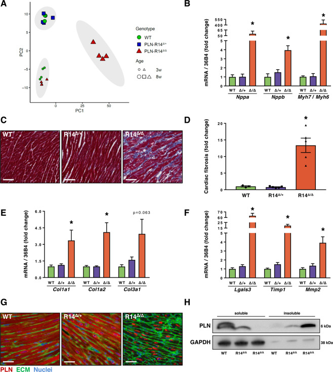
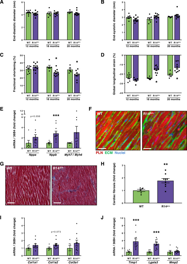
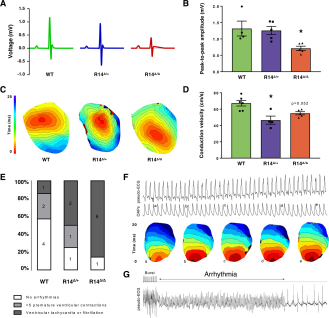
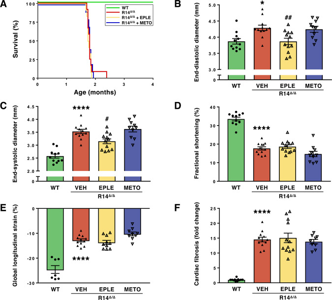
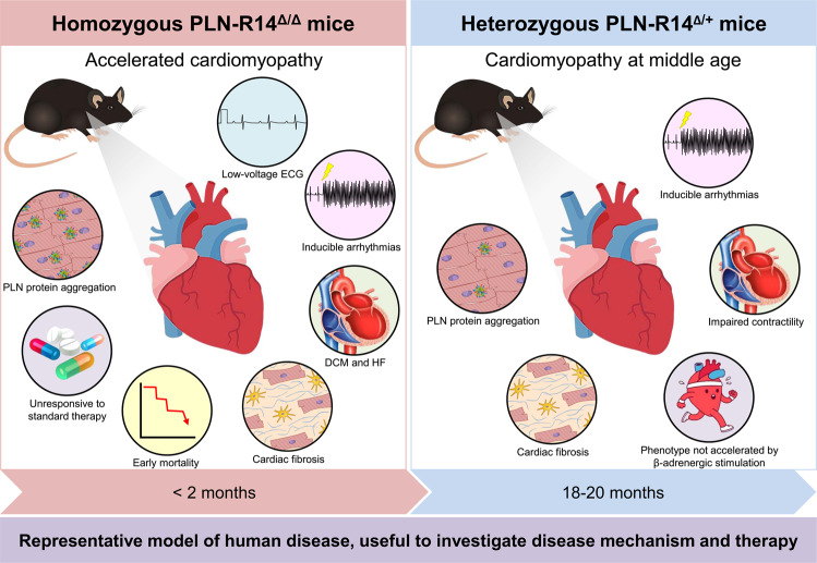
Phospholamban (PLN) plays a role in cardiomyocyte calcium handling as primary inhibitor of sarco/endoplasmic reticulum Ca2+-ATPase (SERCA). The p.(Arg14del) pathogenic variant in the PLN gene results in a high risk of developing dilated or arrhythmogenic cardiomyopathy with heart failure. There is no established treatment other than standard heart failure therapy or heart transplantation. In this study, we generated a novel mouse model with the PLN-R14del pathogenic variant, performed detailed phenotyping, and tested the efficacy of established heart failure therapies eplerenone or metoprolol. Heterozygous PLN-R14del mice demonstrated increased susceptibility to ex vivo induced arrhythmias, and cardiomyopathy at 18 months of age, which was not accelerated by isoproterenol infusion. Homozygous PLN-R14del mice exhibited an accelerated phenotype including cardiac dilatation, contractile dysfunction, decreased ECG potentials, high susceptibility to ex vivo induced arrhythmias, myocardial fibrosis, PLN protein aggregation, and early mortality. Neither eplerenone nor metoprolol administration improved cardiac function or survival. In conclusion, our novel PLN-R14del mouse model exhibits most features of human disease. Administration of standard heart failure therapy did not rescue the phenotype, underscoring the need for better understanding of the pathophysiology of PLN-R14del-associated cardiomyopathy. This model provides a great opportunity to study the pathophysiology, and to screen for potential therapeutic treatments.


Conflict of interest statement

The University Medical Center Groningen, which employs the majority of the authors, has received research grants and/or fees from Abbott, AstraZeneca, Bristol-Myers Squibb, Novartis, Novo Nordisk, and Roche. R.A.d.B. is a minority shareholder of scPharmaceuticals, and received personal fees from Abbott, AstraZeneca, MandalMed. and Novartis. The other authors have no competing interests.

Figures

Figure 1
Figure 1
Generation, validation, survival and cardiac MRI of PLN-R14del mutant mice. (A) Schematic overview of the genomic DNA encoding the murine Pln gene, and the modifications that were performed to replace the WT Pln exon-3 with the R14del Pln exon-3 (marked with 3*). (B) Fluorescent peak trace chromatograms of Sanger sequencing reactions including the coding region of the Pln gene in hearts of WT, PLN-R14Δ/+ and PLN-R14Δ/Δ mice. The codon for the 14th amino acid of the PLN protein is outlined by the grey rectangle. (C) Survival curve of male and female WT, PLN-R14Δ/+ and PLN-R14Δ/Δ mice (n = 6, 10 and 13, respectively). (D) Representative cardiac MRI images at the mid-papillary level in end-diastole and end-systole (scale bar = 1 mm) with quantification of left ventricular end-diastolic volume (E), end-systolic volume (F), stroke volume (G), and ejection fraction (H) of 6-week-old WT, PLN-R14Δ/+ and PLN-R14Δ/Δ mice (n = 4, 5, and 5, respectively). Data are presented as mean ± S.E.M. *p < 0.05 compared to WT (Mann-Whitney test).
Figure 2
Figure 2
Histological and molecular analysis of PLN-R14del mice hearts. (A) Principal component analysis plot of RNA-Seq analysis of left ventricles of WT, PLN-R14Δ/+ and PLN-R14Δ/Δ mice of 3 or 8 weeks of age (n = 4 per group). 95% confidence ellipses of clusters are marked in grey. Information on genes contributing to variance in principle component 1 (PC1, x-axis) and principle component 2 (PC2, y-axis) is shown in Supplementary Fig. 1B. (B) qPCR measurements of left ventricular mRNA levels of Nppa (ANP), Nppb (BNP), and the ratio of Myh7 (β-MHC) to Myh6 (α-MHC) of 8-week-old WT, PLN-R14Δ/+ and PLN-R14Δ/Δ mice (n = 4, 5, and 5, respectively). (C) Representative images of left ventricular sections stained with Masson’s trichrome (scale bar = 70 μm) with (D) quantification of myocardial fibrosis in WT, PLN-R14Δ/+ and PLN-R14Δ/Δ mice hearts (n = 4, 5, and 5, respectively). (E,F) qPCR measurements of left ventricular mRNA levels of cardiac remodelling genes Col1a1, Col1a2, Col3a1, Lgals3 (Gal-3), Timp1, and Mmp2 of 8-week-old WT, PLN-R14Δ/+ and PLN-R14Δ/Δ mice (n = 4, 5, and 5, respectively). (G) Representative images of left ventricular sections of 8-week-old WT, PLN-R14Δ/+ and PLN-R14Δ/Δ mice stained with an anti-PLN antibody (red), wheat-germ agglutinin (WGA) (green), and DAPI (blue) (scale bar = 35 μm). (H) Western blot analysis of monomeric PLN proteins in RIPA-soluble and RIPA-insoluble fractions of left ventricles of 8-week-old WT and PLN-R14Δ/Δ mice (n = 1 and 2, respectively). Images zoom in on the protein bands. Full blot images are presented in Supplementary Fig. S3. Gene expression values are corrected for Rplp0 (36B4) gene expression, and shown as fold change compared to age-matched WT. Myocardial fibrosis is presented as fold change compared to age-matched WT. Data are presented as mean ± S.E.M. *p < 0.05 compared to WT (Mann-Whitney test).
Figure 3
Figure 3
Cardiac functional, histological and molecular analysis of 12- to 20-month-old PLN-R14Δ/+ mice. Echocardiographic analysis of left ventricular end-diastolic diameter (A), end-systolic diameter (B), fractional shortening (C), and global longitudinal strain (D) of 12-, 18- and 20-month-old WT and PLN-R14Δ/+ mice (n = 9, 10, 6, 10, 5 and 9, respectively). (E) qPCR measurements of left ventricular mRNA levels of Nppa (ANP), Nppb (BNP), and the ratio of Myh7 (β-MHC) to Myh6 (α-MHC) of 20-month-old WT and PLN-R14Δ/+ mice (n = 6 and 10, respectively). (F) Representative images of left ventricular sections of 20-month-old WT and PLN-R14Δ/+ mice stained with an anti-PLN antibody (red), wheat-germ agglutinin (WGA) (green), and DAPI (blue) (scale bar = 35 μm) or (G) stained with Masson’s trichrome (scale bar = 70 μm) with (H) quantification of myocardial fibrosis (n = 6 and 9, respectively). (I,J) qPCR measurements of left ventricular mRNA levels of cardiac remodelling genes Col1a1, Col1a2, Col3a1, Lgals3 (Gal-3), Timp1, and Mmp2 of 20-month-old WT and PLN-R14Δ/+ mice (n = 6 and 10, respectively). Gene expression values are corrected for Rplp0 (36B4) gene expression, and shown as fold change compared to age-matched WT. Myocardial fibrosis is presented as fold change compared to age-matched WT. Data are presented as mean ± S.E.M. *p < 0.05, **p < 0.01, ***p < 0.001 compared to age-matched WT (Mann-Whitney test).
Figure 4
Figure 4
Electrophysiological characterization of PLN-R14del mice hearts. (A) Representative averaged views of 1-minute in vivo ECG measurements of 6-week-old WT, PLN-R14Δ/+ and PLN-R14Δ/Δ mice with (B) quantification of total QRS-complex (peak-to-peak) amplitude (n = 4, 5, and 5, respectively). (C) Reconstructed activation maps during ex vivo left ventricular stimulation (120 ms interval) with (D) quantification of longitudinal conduction velocity in WT, PLN-R14Δ/+ and PLN-R14Δ/Δ mice hearts (n = 6, 4, and 5, respectively). (E) Incidence of ex vivo induced ventricular arrhythmias in WT, PLN-R14Δ/+ and PLN-R14Δ/Δ mice hearts (n = 7, 4, and 7, respectively). (F) Example of a pseudo-ECG (upper) with simultaneously recorded optical action potentials (OAPs) (middle), and reconstructed activation maps (lower) from a WT mouse, which indicates the order of activation from several beats that correspond to the traces above (indicated by the small letter). (G) Pseudo-ECG showing an induced arrhythmia in a PLN-R14Δ/Δ mouse heart following burst pacing. Data are presented as mean ± S.E.M. *p < 0.05 compared to WT (Mann-Whitney test).
Figure 5
Figure 5
Effect of administration of eplerenone or metoprolol on survival and cardiac function of PLN-R14Δ/Δ mice. (A) Survival curve of WT and PLN-R14Δ/Δ mice without or with eplerenone (EPLE; 200 mg/kg/day) or metoprolol (METO; 350 mg/kg/day) administration (n = 11, 12, 12, and 10, respectively). (B–E) Echocardiographic analysis of left ventricular end-diastolic and end-systolic diameter, fractional shortening, and global longitudinal strain of 6-week-old WT and PLN-R14Δ/Δ mice without or with eplerenone or metoprolol administration (n = 11, 12, 12, and 10, respectively). (F) Quantification of myocardial fibrosis in Masson’s trichrome-stained left ventricular sections of 16-week-old WT and 8-week-old PLN-R14Δ/Δ mice without or with eplerenone or metoprolol administration (n = 11, 12, 12, and 10, respectively). Myocardial fibrosis is presented as fold change compared to age-matched WT. Data are presented as mean ± S.E.M. *p < 0.05, ****p < 0.0001 compared to WT; #p < 0.05, ##p < 0.01 compared to R14Δ/Δ + VEH (one-way ANOVA followed by Tukey’s post-hoc test).
Figure 6
Figure 6
Graphical abstract. PLN-R14Δ/Δ mice (left) mimic human disease in a strikingly comparable but accelerated manner, evidenced by cardiac dilatation, contractile dysfunction, decreased ECG potentials, high susceptibility to ex vivo induced arrhythmias, cardiac fibrosis, PLN protein aggregation, and early mortality. Administration of standard HF therapy could not rescue the phenotype. PLN-R14Δ/+ mice (right) demonstrated increased susceptibility to ex vivo induced arrhythmias, and developed cardiomyopathy with comparable characteristics at middle age, similar to human carriers. The phenotype was not accelerated by β-adrenergic stimulation.

References

    1. MacLennan DH, Kranias EG. Phospholamban: a crucial regulator of cardiac contractility. Nat. Rev. Mol. Cell Biol. 2003;4:566–577. - PubMed
    1. Fish M, et al. Mutation analysis of the phospholamban gene in 315 South Africans with dilated, hypertrophic, peripartum and arrhythmogenic right ventricular cardiomyopathies. Sci. Rep. 2016;6:22235. - PMC - PubMed
    1. Haghighi K, et al. A mutation in the human phospholamban gene, deleting arginine 14, results in lethal, hereditary cardiomyopathy. Proc. Natl. Acad. Sci. 2006;103:1388–1393. - PMC - PubMed
    1. DeWitt MM, MacLeod HM, Soliven B, McNally EM. Phospholamban R14 deletion results in late-onset, mild, hereditary dilated cardiomyopathy. J. Am. Coll. Cardiol. 2006;48:1396–1398. - PubMed
    1. Cheung CC, et al. Phospholamban cardiomyopathy: a Canadian perspective on a unique population. Netherlands Hear. J. 2019;27:208–213. - PMC - PubMed

Publication types