

GlioM&M: Web-based tool for studying circulating and infiltrating monocytes and macrophages in glioma
- PMID: 32555306
- PMCID: PMC7303151
- DOI: 10.1038/s41598-020-66728-w
GlioM&M: Web-based tool for studying circulating and infiltrating monocytes and macrophages in glioma
Abstract
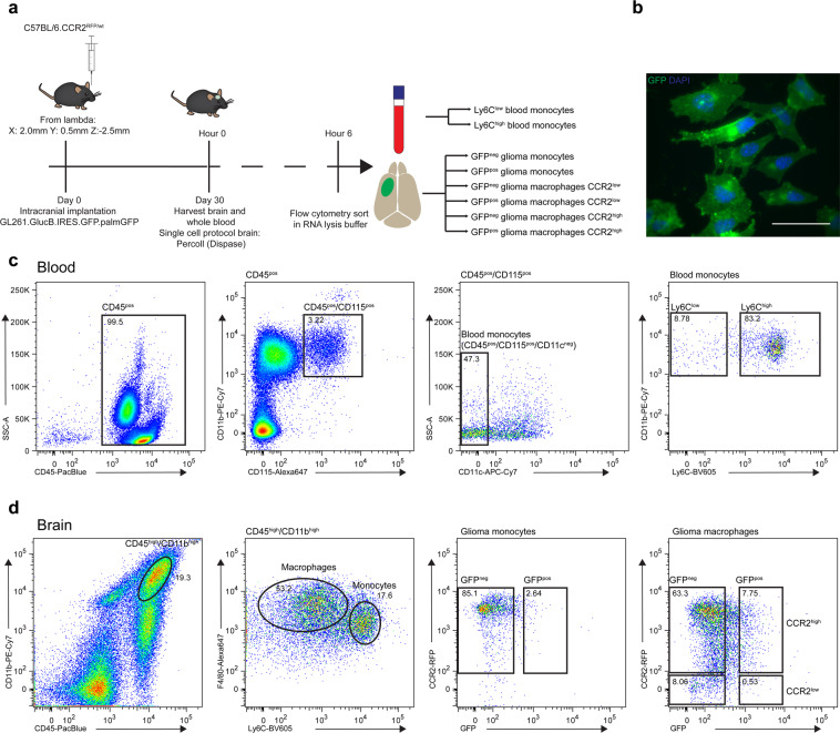
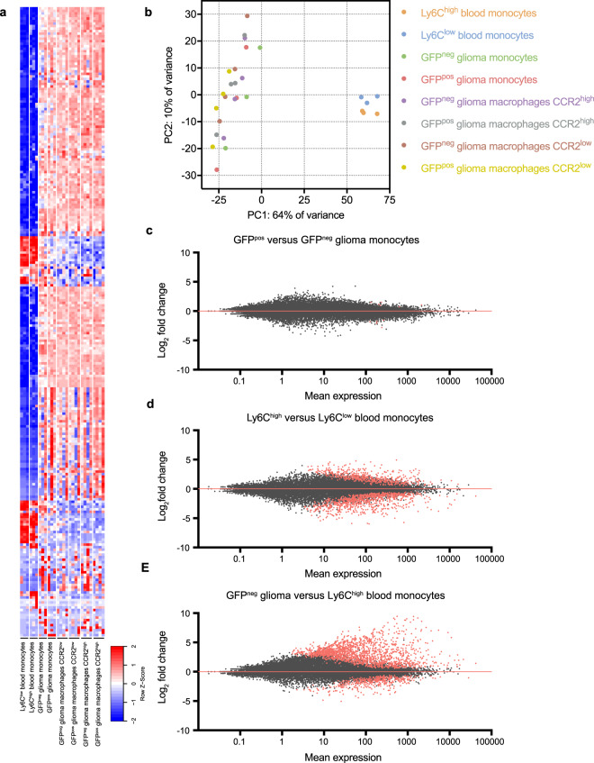
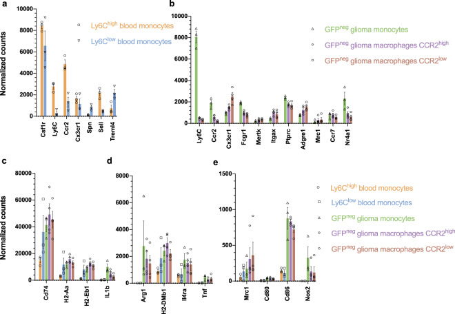
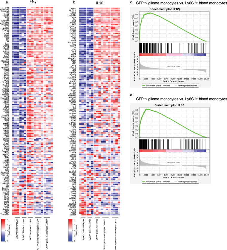
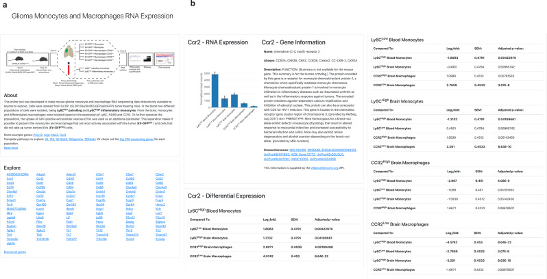
Monocytes, macrophages and microglia make up a large part of the glioma environment and have an important role in maintaining and propagating glioma progression. Targeting these cells to inhibit their tumor-promoting effect and reprogramming them into an anti-tumor phenotype is a potential therapeutic approach for glioma. In this study we analyzed the transcriptomes of eight different monocyte subgroups derived from the brain and the blood of glioma-bearing mice. We compared the expression profile of blood-derived monocytes versus tumor-infiltrating monocytes and found increased expression of both pro- and anti-inflammatory pathways in tumor infiltrating monocytes. To help disseminate these datasets, we created a user-friendly web-based tool accessible at www.glioma-monocytes.com. This tool can be used for validation purposes and to elucidate gene expression profiles of tumor-interacting monocytes and macrophages as well as blood-derived circulating monocytes. This tool can also be used to identify new markers and targets for therapy in these different cell populations.
Conflict of interest statement
The authors declare no competing interests.
Figures
Similar articles
-
Tumor-infiltrating myeloid-derived suppressor cells are pleiotropic-inflamed monocytes/macrophages that bear M1- and M2-type characteristics.J Leukoc Biol. 2008 May;83(5):1136-44. doi: 10.1189/jlb.0907611. Epub 2008 Feb 19. J Leukoc Biol. 2008. PMID: 18285406
-
Deletion of the RNA regulator HuR in tumor-associated microglia and macrophages stimulates anti-tumor immunity and attenuates glioma growth.Glia. 2019 Dec;67(12):2424-2439. doi: 10.1002/glia.23696. Epub 2019 Aug 10. Glia. 2019. PMID: 31400163 Free PMC article.
-
Distinct roles of CSF family cytokines in macrophage infiltration and activation in glioma progression and injury response.J Pathol. 2013 Jul;230(3):310-21. doi: 10.1002/path.4192. J Pathol. 2013. PMID: 23520016
-
When Immune Cells Turn Bad-Tumor-Associated Microglia/Macrophages in Glioma.Int J Mol Sci. 2018 Feb 1;19(2):436. doi: 10.3390/ijms19020436. Int J Mol Sci. 2018. PMID: 29389898 Free PMC article. Review.
-
Microglia/Brain Macrophages as Central Drivers of Brain Tumor Pathobiology.Neuron. 2019 Nov 6;104(3):442-449. doi: 10.1016/j.neuron.2019.08.028. Neuron. 2019. PMID: 31697921 Free PMC article. Review.
Cited by
-
Radio-chemotherapy and metformin selectively modulate the heterogeneous landscape of glioma with ribosome biogenesis, long non coding RNA and immune-escape markers as major player.Int J Biol Sci. 2025 May 27;21(8):3527-3554. doi: 10.7150/ijbs.103194. eCollection 2025. Int J Biol Sci. 2025. PMID: 40520013 Free PMC article.
-
Pro- vs. Anti-Inflammatory Features of Monocyte Subsets in Glioma Patients.Int J Mol Sci. 2023 Jan 18;24(3):1879. doi: 10.3390/ijms24031879. Int J Mol Sci. 2023. PMID: 36768201 Free PMC article.
-
Nanotechnology-Based Combinatorial Anti-Glioblastoma Therapies: Moving from Terminal to Treatable.Pharmaceutics. 2022 Aug 15;14(8):1697. doi: 10.3390/pharmaceutics14081697. Pharmaceutics. 2022. PMID: 36015322 Free PMC article. Review.
-
Targeting the Endocannabinoid System Present in the Glioblastoma Tumour Microenvironment as a Potential Anti-Cancer Strategy.Int J Mol Sci. 2024 Jan 23;25(3):1371. doi: 10.3390/ijms25031371. Int J Mol Sci. 2024. PMID: 38338649 Free PMC article. Review.
-
The Prognostic Impact of Gender, Therapeutic Strategies, Molecular Background, and Tumor-Infiltrating Lymphocytes in Glioblastoma: A Still Unsolved Jigsaw.Genes (Basel). 2023 Feb 15;14(2):501. doi: 10.3390/genes14020501. Genes (Basel). 2023. PMID: 36833428 Free PMC article.
References
Publication types
MeSH terms
Substances
Grants and funding
LinkOut - more resources
Full Text Sources
Medical
Molecular Biology Databases
