. 2020 Jun 18;10(1):9914.
doi: 10.1038/s41598-020-66020-x.

Molecular approaches underlying the oogenic cycle of the scleractinian coral, Acropora tenuis

Affiliations

Molecular approaches underlying the oogenic cycle of the scleractinian coral, Acropora tenuis

Ee Suan Tan et al. Sci Rep. .

Abstract

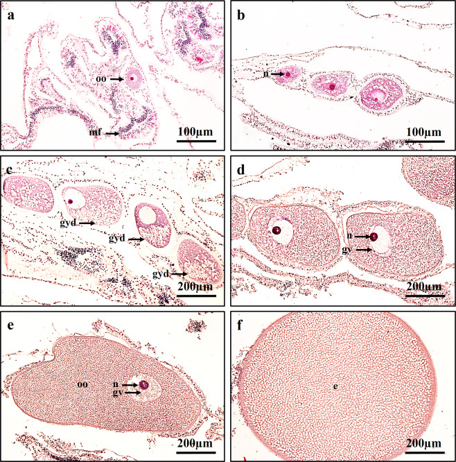
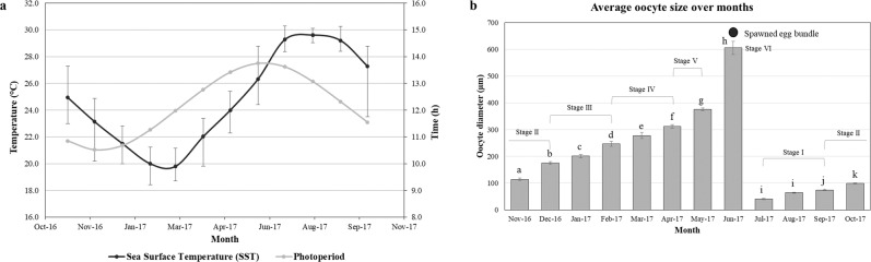
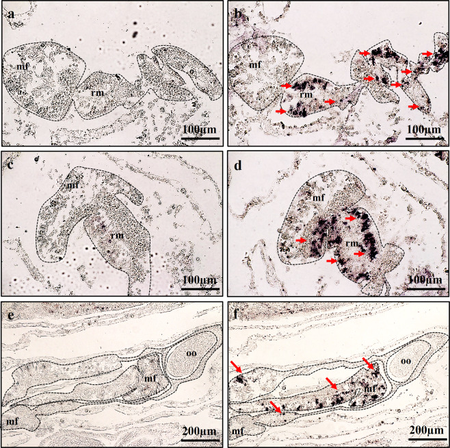
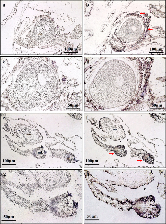
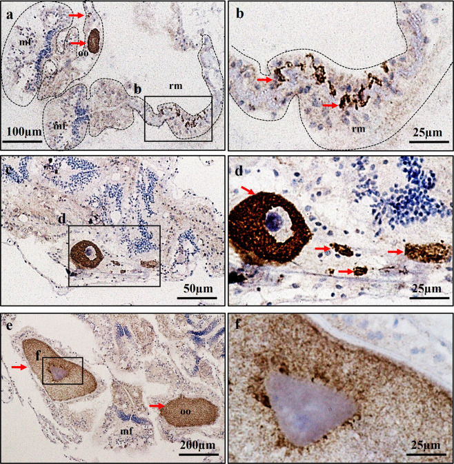
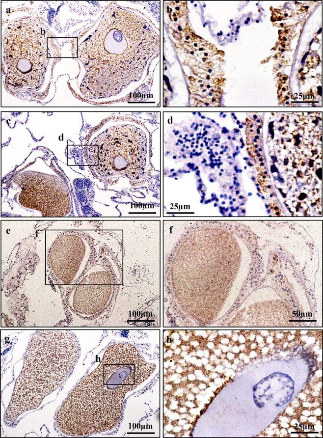
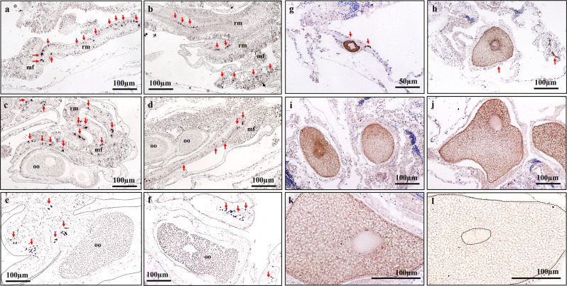
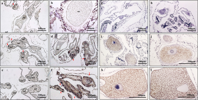
This study aimed to elucidate the physiological processes of oogenesis in Acropora tenuis. Genes/proteins related to oogenesis were investigated: Vasa, a germ cell marker, vitellogenin (VG), a major yolk protein precursor, and its receptor (LDLR). Coral branches were collected monthly from coral reefs around Sesoko Island (Okinawa, Japan) for histological observation by in situ hybridisation (ISH) of the Vasa (AtVasa) and Low Density Lipoprotein Receptor (AtLDLR) genes and immunohistochemistry (IHC) of AtVasa and AtVG. AtVasa immunoreactivity was detected in germline cells and ooplasm, whereas AtVG immunoreactivity was detected in ooplasm and putative ovarian tissues. AtVasa was localised in germline cells located in the retractor muscles of the mesentery, whereas AtLDLR was localised in the putative ovarian and mesentery tissues. AtLDLR was detected in coral tissues during the vitellogenic phase, whereas AtVG immunoreactivity was found in primary oocytes. Germline cells expressing AtVasa are present throughout the year. In conclusion, Vasa has physiological and molecular roles throughout the oogenic cycle, as it determines gonadal germline cells and ensures normal oocyte development, whereas the roles of VG and LDLR are limited to the vitellogenic stages because they act in coordination with lipoprotein transport, vitellogenin synthesis, and yolk incorporation into oocytes.


Conflict of interest statement

The authors declare no competing interests.

Figures

Figure 1
Figure 1
Distinct oocyte stages of A. tenuis. (a) Stage I; oocyte enveloped in mesoglea, showing nucleus. (b) Stage II; oocyte, cytoplasm, and nucleus have grown in size. (c) Stage III; oocyte with finely granular yolk/cytoplasm. Yolk polarity and migration of nucleolus to periphery of nucleus observed. (d,e) Stage IV-V; oocyte grow tremendously in size. (f) Stage VI; Final stage (Egg). oo; oocyte, mf; mesentery filament, n; nucleus, gv; germinal vesicle, gyd; granular yolk disposition, e; egg.
Figure 2
Figure 2
(a) Annual changes in average sea surface temperature and photoperiod in Sesoko station, Okinawa, Japan. Black line indicates Sea Surface Temperature (SST) and grey line indicates Photoperiod. Error bar indicates maximum and minimum value of SST. (b) Monthly changes in mean diameter of oocytes of A. tenuis. The data are presented as mean ± SE. Significant differences (P < 0.05) are indicated with the different lower case letters. Respective oocyte stages are shown above each bar graphs.
Figure 3
Figure 3
Expression profiles of AtVasa transcript in mesentery tissues. (a–f) Localization of AtVAsa mRNA-positive cells in the retractor muscles (rm) area of mesentery tissues. (a,c,e) showed section stained with AtVasa sense probe. (b,d,f) showed section stained with AtVasa anti-sense probe. Purple colouration (ALP reaction) indicates AtVasa transcript signals. mf; mesenterial filament, oo; oocyte.
Figure 4
Figure 4
Expression profiles of AtLDLR transcript in putative ovarian and mesentery tissues. (a–h) Localization of AtLDLR mRNA-positive cells in putative ovarian and mesentery tissue. (a,c,e,g) showed section stained with AtLDLR sense probe. (b,d,f,h) showed section stained with AtLDLR anti-sense probe. Purple colouration (ALP reaction) indicates AtLDLR transcript signals. (c,d,g,h) showed higher magnification views of (a,b,e,f) respectively. mf; mesenterial filament, oo; oocyte.
Figure 5
Figure 5
Characterization of Vasa immunoreactivity (AtVasa) in A. tenuis tissues. (a) AtVasa positive cells located in germ cells in the retractor muscle (rm); (b) A higher magnification view of the inset shown in (a). (c) AtVasa positive early oocyte and germ cells located along the meseoglea; (d) A higher magnification view of inset shown in (c). (e) AtVasa positive in oocyte cytoplasm; (f) A higher magnification view of inset shown in (e) Arrow indicates AtVasa immunoreactivity signals. mf; mesenterial filament, oo; oocyte.
Figure 6
Figure 6
Characterization of VG immunoreactivity (AtVG) in A. tenuis tissues. (a,c) AtVG positive signal located in putative ovarian tissues; (b,d) A higher magnification view of the inset shown in (a,c) respectively; (e,g) AtVG positive in oocyte cytoplasm; (f,h) A higher magnification view of inset shown in (e,g) respectively. Arrow indicates AtVG immunoreactivity signals.
Figure 7
Figure 7
The presence of mRNA expression of AtVasa in germ cells (a–f) and immunoreactivity of AtVasa (g–l) in A. tenuis oocytes at different development stages of oogenesis. ISH: (a,b) Stage I-II; (c,d) Stage III; (d,e) Stage IV-V. IHC: (g) Stage I; (h) Stage II; (i) Stage III; (j,k) Stage IV; (l) Stage V. Arrow indicate AtVasa transcript and AtVasa immunoreactivity. rm; retractor muscle, mf; mesentery filament, oo; oocyte.
Figure 8
Figure 8
Comparison of mRNA expression of AtLDLR during non-vitellogenic phase, Stage I & II (a,c,e) and vitellogenic phase, Stage III-V (b,d,f) of A. tenuis. Meanwhile. (g-l) shows the seasonal change of AtVG immunoreactivity at different development stages of oogenesis. IHC: (g). Stage I; (h) Stage II; (i–j) Stage III; (k) Stage IV; (l) Stage V. oo; oocyte. Arrow indicate AtLDLR transcript.h.

References

    1. Chui PY, et al. Gametogenesis, Embryogenesis, and Fertilization Ecology of Platygyra acuta in Marginal Nonreefal Coral Communities in Hong Kong. J. Mar. Biol. 2014;2014:1–9.
    1. Harrison, P. L. Sexual reproduction in scleractinian corals in Coral reefs: an ecosystem in transition (eds. Dubinsky, Z., Stambler, N.) 59-85 (Springer, Dordrecht, 2011).
    1. Harrison, P. L., Wallace, C. C. Reproduction, dispersal and recruitment of scleractinian coral in Ecosystem of the World Vol. 25 (ed. Dubinsky, Z.) 133–207 (Elsevier, Amsterdam, 1990).
    1. Isomura, N. & Fukami, H. Coral Reproduction in Japan in Coral Reef Studies of Japan (ed. Iguchi A.) 95–110 (Springer, Dordrecht, 2018).
    1. Harrison PL, et al. Mass spawning in tropical reef corals. Science. 1984;223:1186–1189. - PubMed

Publication types