

doi: 10.1038/s41423-020-0485-9.
Epub 2020 Jun 18.
The ORF3a protein of SARS-CoV-2 induces apoptosis in cells
Affiliations
- PMID: 32555321
- PMCID: PMC7301057
- DOI: 10.1038/s41423-020-0485-9
Item in Clipboard
The ORF3a protein of SARS-CoV-2 induces apoptosis in cells
Cell Mol Immunol.
2020 Aug.
No abstract available
Conflict of interest statement
The authors declare no competing interests.
Figures
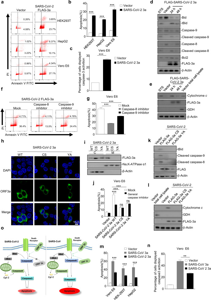
a, b HEK293T, HepG2, and Vero E6 cells were transfected with FLAG-SARS-CoV-2 ORF3a. After 24 h, cells were stained with annexin V-fluorescein 5-isothiocyanate (FITC)/propidium iodide (PI) for flow cytometric analysis (a), and the percentage of apoptotic cells was measured (b). c Vero E6 cells were transfected with FLAG-SARS-CoV-2 ORF3a. After 24 h, cells were stained with caspase-3/7 green detection reagent for fluorescence analysis, and the percentage of cells displaying caspase-3 activation was measured. d HEK293T cells were transfected with empty vector or FLAG-SARS-CoV-2 ORF3a. After 12 and 24 h, cells were subjected to western blotting analysis using the indicated antibodies. Cells treated with STS for 5 h were used as a positive control. STS, staurosporine. e HEK293T cells transfected with empty vector or FLAG-SARS-CoV-2 ORF3a for 12 and 24 h, or cells treated with STS for 5 h, were collected and the mitochondria were separated via gradient centrifugation. Cell lysates without mitochondria were subjected to western blotting using the indicated antibodies. The total cell lysates within intact mitochondria were used as positive control. GDH, glutamate dehydrogenase. f, g Vero E6 cells were transfected with FLAG-SARS-CoV-2 ORF3a in the presence of DMSO, caspase-8 inhibitor, or caspase-9 inhibitor. After 24 h, cells were stained with annexin V-FITC/PI for flow cytometric analysis (f), and the percentage of apoptotic cells was measured (g). h HEK293T cells were transfected with FLAG-SARS-CoV-2 ORF3a and its mutants (CS and YA). After 24 h, cells were stained with a mouse anti-FLAG antibody and Alexa-488 conjugated anti-mouse IgG for immunofluorescence. Scale bar, 10 μM. i HEK293T cells were transfected with FLAG-SARS-CoV-2 ORF3a and its mutants (CS and YA). After 24 h, cells were collected and the membrane and plasma proteins were separately extracted for western blotting. j Vero E6 cells were transfected with FLAG-SARS-CoV-2 ORF3a mutants (CS and YA) and treated with DMSO or a general caspase inhibitor. After 24 h, cells were stained with annexin V-FITC/PI for flow cytometric analysis, and the percentage of apoptotic cells was measured. k, l HEK293T cells were transfected with vector or FLAG-SARS-CoV-2 ORF3a and its mutants (CS and YA). After 24 h, cells were collected and the membrane and plasma proteins were separately extracted for western blotting (k). To examine levels of cytochrome c in the cytosol, mitochondria were separated via gradient centrifugation, and cell lysates without mitochondria were subjected to western blotting (l). m HEK293T, HepG2, and Vero E6 cells were transfected with vector, FLAG-SARS-CoV ORF3a, or FLAG-SARS-CoV-2 ORF3a. After 24 h, cells were stained with annexin V-FITC/PI for flow cytometric analysis, and the percentage of apoptotic cells was measured. n Vero E6 cells were transfected with vector, FLAG-SARS-CoV ORF3a, or FLAG-SARS-CoV-2 ORF3a. After 24 h, caspase-3 activation was detected by using caspase-3/7 green detection reagent, and the percentage of cells displaying caspase-3 activation was measured. o Left, the pro-apoptotic activity of SARS-CoV-2 ORF3a requires the membrane association of ORF3a. Right, membrane association is involved but not essential for the pro-apoptotic activity of SARS-CoV ORF3a, and SARS-CoV ORF3a can induce apoptosis in a membrane-independent manner. SARS-CoV-2 ORF induces apoptosis in a lesser extent than that of SARS-CoV ORF3a. **p < 0.01, ***p < 0.001 by two-tailed Student’s t test
References
-
- Qin L, Jeng H, Rakue Y, Mizota T. A deficient public health system as a contributing cause of Severe Acute Respiratory Syndrome (SARS) epidemic in mainland China. Southeast Asian J. Trop. Med. Public Health. 2005;36:213–216. - PubMed
Publication types
MeSH terms
Substances
LinkOut - more resources
Full Text Sources
Other Literature Sources
Research Materials
Miscellaneous
