Review
. 2020 Jul;19(7):480-494.
doi: 10.1038/s41573-020-0070-z. Epub 2020 Jun 17.

Targeting the Hippo pathway in cancer, fibrosis, wound healing and regenerative medicine

Affiliations
Review

Targeting the Hippo pathway in cancer, fibrosis, wound healing and regenerative medicine

Anwesha Dey et al. Nat Rev Drug Discov. 2020 Jul.

Abstract

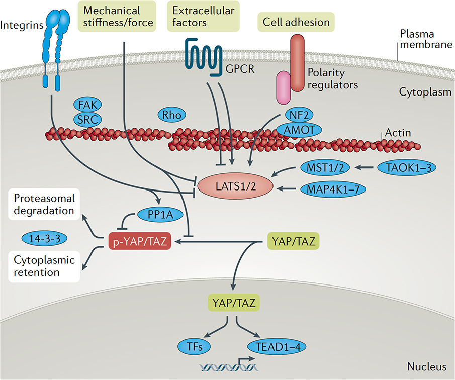
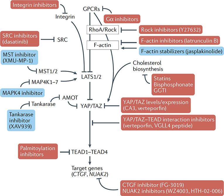
The Hippo pathway is an evolutionarily conserved signalling pathway with key roles in organ development, epithelial homeostasis, tissue regeneration, wound healing and immune modulation. Many of these roles are mediated by the transcriptional effectors YAP and TAZ, which direct gene expression via control of the TEAD family of transcription factors. Dysregulated Hippo pathway and YAP/TAZ-TEAD activity is associated with various diseases, most notably cancer, making this pathway an attractive target for therapeutic intervention. This Review highlights the key findings from studies of Hippo pathway signalling across biological processes and diseases, and discusses new strategies and therapeutic implications of targeting this pathway.


Conflict of interest statement

Competing interests

K.-L.G. is a co-founder and has an equity interest in Vivace Therapeutics, Inc. The terms of this arrangement have been reviewed and approved by the University of California, San Diego in accordance with its conflict-of-interest policies. A.D. is an employee of Genentech and shareholder at Roche.

Figures

Fig 1.
Fig 1.. Key signals regulating YAP/TAZ activity.
YAP and TAZ activity is regulated by the LATS1 and LATS2 kinases, which phosphorylate YAP/TAZ on conserved residues. Phosphorylated YAP/TAZ (p-YAP/TAZ) associate with 14-3-3 proteins and are retained in the cytoplasm and targeted for degradation via the proteasome, consequently leading to low nuclear levels. Various upstream effectors of the LATS1 and LATS2 kinases have been identified, including the MST, MAP4K and TAOK families of kinases, which phosphorylate and activate LATS1/2. Cell polarity and adhesion regulators facilitate altered actin dynamics and Hippo pathway effector association to promote LATS1/2-mediated regulation of YAP/TAZ. G protein-coupled receptors (GPCRs), mechanical cues and signals transduced by the extracellular matrix and matrix-binding integrins - such as those transduced by FAK and SRC - can inactivate LATS1/2 or induce the dephosphorylation of YAP/TAZ via PP1A activation, collectively leading to hypo-phosphorylated YAP/TAZ. Hypo-phosphorylated YAP and TAZ accumulate in the nucleus, where they can bind to various transcription factors (TFs), most notably the TEAD family, to direct gene expression changes that control a range of biological events.
Fig. 2.
Fig. 2.. Proposed strategies and small molecules (direct and indirect) that could promote or inhibit YAP/TAZ activity.
Opportunities exist for small molecules for biological intervention of key YAP/TAZ-regulatory signals that may offer targeted medical promise. These include various targets that would result in repression of YAP/TAZ nuclear activity (shown in red boxes), such as inhibitors of repressors of LATS1/2 activity, inhibitors of YAP/TAZ interacting transcription factors (such as the TEAD family) or inhibitors of important downstream target genes that drive aberrant biological processes. Activation of YAP/TAZ may also be desirable, such as in the context of medical regenerative strategies, and may be achieved by repressing the activity of effectors that promote YAP/TAZ cytoplasmic retention (shown in blue boxes). CTGF, connective tissue growth factor; GPCR, G protein-coupled receptor.
Fig. 3.
Fig. 3.. Structural biology of the TEAD–YAP interface and the TEAD lipid pocket.
(a) TEAD proteins contain a highly conserved carboxy-terminal YAP/TAZ binding domain (YBD). The binding interface between the TEAD YBD (blue) and the TEAD-binding peptide of YAP (teal) comprises three highly conserved interfaces (sites 1, 2 and 3) that contribute to high-affinity binding. TEADs also contain a central hydrophobic lipid pocket (shown at the right) with an Spalmitoyl cysteine (yellow sticks) modification at a universally conserved cysteine. (b) Snapshots of the lipid pockets of all four TEADs highlight the difficulty in developing pan-TEAD lipid pocket ligands as well as providing a rubric for the design of isoform-specific ligands. Residues shown are lipid pocket residues that are nonconserved in at least one of the four human TEAD paralogues. (c) Schematic representation of the human TEAD lipid pocket. Residues in black as well as those highlighted in red are invariant among human TEADs (residues in red are directly important for lipidation). Those highlighted in blue are non-identical in at least one human TEAD paralogue but still similar. Residues highlighted in pink are non-identical and non-similar in at least one paralogue.
Fig. 4.
Fig. 4.. Representative compounds targeting the Hippo pathway.
Key potentially druggable sites in the protein–protein interaction between YAP/TAZ and TEAD have been identified, as well as a highly conserved palmitoylation pocket in TEADs. Some small molecules that target these interactions are shown, such as Peptide 17 ,. Although compounds such as verteporfin or CA3 successfully inhibit TEAD transcriptional activity and have shown antitumour activity in vivo, the target specificity and selectivity of these compounds remain to be confirmed. Other approaches have focused on disrupting the interaction between TEAD and other transcriptional co-activators, such as vestigial-like protein family members (VGLL1-VGLL4) with VGLL4-mimmicking peptides, such as ‘super-TDU’. Finally, compounds such as flufenamic acid and TED-347, which target the lipid pocket at the core of all four TEADs and inhibit palmitoylation, have shown antitumour activity in vitro. TDU, Tondu.

References

    1. Jia J, Zhang W, Wang B, Trinko R & Jiang J The Drosophila Ste20 family kinase dMST functions as a tumor suppressor by restricting cell proliferation and promoting apoptosis. Genes Dev. 17, 2514–2519 (2003). - PMC - PubMed
    1. Justice RW, Zilian O, Woods DF, Noll M & Bryant PJ The Drosophila tumor suppressor gene Warts encodes a homolog of human myotonic dystrophy kinase and is required for the control of cell shape and proliferation. Genes Dev. 9, 534–546 (1995). - PubMed
    1. Kango-Singh M et al. Shar-pei mediates cell proliferation arrest during imaginal disc growth in Drosophila. Development 129, 5719–5730 (2002). - PubMed
    1. Pantalacci S, Tapon N & Leopold P The Salvador partner Hippo promotes apoptosis and cell-cycle exit in Drosophila. Nat. Cell Biol. 5, 921–927 (2003). - PubMed
    1. Tapon N et al. Salvador promotes both cell cycle exit and apoptosis in Drosophila and is mutated in human cancer cell lines. Cell 110, 467–478 (2002). - PubMed

MeSH terms