Review
. 2020 Nov;17(11):732-747.
doi: 10.1038/s41569-020-0394-8. Epub 2020 Jun 18.

Intracellular calcium leak in heart failure and atrial fibrillation: a unifying mechanism and therapeutic target

Affiliations
Review

Intracellular calcium leak in heart failure and atrial fibrillation: a unifying mechanism and therapeutic target

Haikel Dridi et al. Nat Rev Cardiol. 2020 Nov.

Abstract

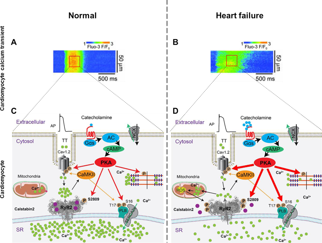
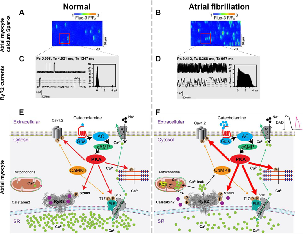
Ca2+ is a fundamental second messenger in all cell types and is required for numerous essential cellular functions, including cardiac and skeletal muscle contraction. The intracellular concentration of free Ca2+ ([Ca2+]) is regulated primarily by ion channels, pumps (ATPases), exchangers and Ca2+-binding proteins. Defective regulation of [Ca2+] is found in a diverse spectrum of pathological states that affect all the major organs. In the heart, abnormalities in the regulation of cytosolic and mitochondrial [Ca2+] occur in heart failure (HF) and atrial fibrillation (AF), two common forms of heart disease and leading contributors to morbidity and mortality. In this Review, we focus on the mechanisms that regulate ryanodine receptor 2 (RYR2), the major sarcoplasmic reticulum (SR) Ca2+-release channel in the heart, how RYR2 becomes dysfunctional in HF and AF, and its potential as a therapeutic target. Inherited RYR2 mutations and/or stress-induced phosphorylation and oxidation of the protein destabilize the closed state of the channel, resulting in a pathological diastolic Ca2+ leak from the SR that both triggers arrhythmias and impairs contractility. On the basis of our increased understanding of SR Ca2+ leak as a shared Ca2+-dependent pathological mechanism in HF and AF, a new class of drugs developed in our laboratory, known as rycals, which stabilize RYR2 channels and prevent Ca2+ leak from the SR, are undergoing investigation in clinical trials.


Figures

Figure 1.
Figure 1.. Diastolic SR Ca2+ leak in ventricular myocytes from failing hearts.
Representative line scan of Ca2+ sparks recorded in ventricular cardiomyocytes from normal (A) and failing murine hearts (B). Representative RyR2 single-channel tracings under conditions that simulate diastole when the cytosolic Ca2+ concentration is low (e.g. 100–150 nM), in normal (C) and heart failure (D) cardiomyocytes . Schematic representation of intracellular Ca2+ release in EC coupling in normal (E) and heart failure (F) cardiomyocytes. There is little or no diastolic SR Ca2+ leak in ventricular cardiomyocytes from normal non-failing hearts (panels A,C and E). In contrast, ventricular cardiomyocytes from failing hearts exhibit increased spontaneous Ca2+ sparks, increased RyR2 open probability, diastolic SR Ca2+ leak and reduced SR Ca2+ stores, all consistent with a pathologic leak of SR Ca2+via RyR2 remodeled (including PKA hyperphosphorylation, oxidation, nitrosylation and dissociation of calstabin2 from the RyR2 channel complex) resulting in RyR2 channels that do not close properly during diastole. (panels B,D and F). This results in reduced SR Ca2+ content. AP, action potential; TT, transverse tubule; cAMP, cyclic adenosine monophosphate; Cav1.2, L-type Ca2+ channel; NCX, Na+/Ca2+ exchanger; RyR2, ryanodine receptor type-2; SERCA2a, sarco/endoplasmic reticulum ATPase type-2a; PLB, phospholamban; PKA, protein kinase A; CaMKII, Ca2+/calmodulin-dependent protein kinase II; ROS, reactive oxygen species.
Figure 2.
Figure 2.. Reduced Ca2+ transients in ventricular myocytes from failing hearts.
Representative line scan showing Ca2+ waves (transients) recorded in ventricular cardiomyocytes from normal (A) and failing hearts (B). The Ca2+ transient amplitude is lower in HF ventricular cardiomyocytes primarily because of the reduced SR Ca2+ content due to RyR2 diastolic leak and reduced SERCA2a, and the Ca2+ transient duration is longer primarily due to slower re-uptake kinetics compared with normal ventricular cardiomyocytes resulting in impaired contractility in failing hearts. Schematic representation of Ca2+ signaling in normal (C) and heart failure (D) ventricular myocytes. Of note RyR1 in skeletal muscle are also leaky during HF. This contributes to impaired exercise capacity which is a common symptom in patients with advanced (Stage III and IV) HF .
Figure 3.
Figure 3.. SR Ca2+ leak triggers AF in atrial myocytes.
Representative line scan of Ca2+ sparks recorded in atrial myocytes from normal (A) and atrial fibrillation (B). Representative RyR2 single-channel tracings recorded under conditions simulating diastole in atrial cardiomyocytes at a resting [Ca2+]cyt ≃ 150 nM, in normal (C) and atrial fibrillation cardiomyocytes (D). Schematic representation of Ca2+ signaling in normal (E) and atrial fibrillation (F) atrial myocytes. Atrial myocyte exhibit increased spontaneous Ca2+ sparks, increased RyR2 open probability, Ca2+ leak and reduced SR Ca2+ stores in AF.
Figure 4.
Figure 4.. RyR2 phosphorylation domain and CPVT-associated mutation hot spots.
CPVT-associated RyR2 mutations (http://www.uniprot.org/uniprot/Q92736) in the N-terminal domain (red spheres), bridging solenoid (blue spheres), core solenoid (purple spheres), transmembrane domain (orange spheres), and C-terminal domain (magenta), viewed from the cytosol (left) and from the plane of the SR membrane (right). Insets show the domain-domain interface between the NTD and the bridging solenoid at the ‘zipping’ point where DPc10 (black ribbon) is located, the newly revealed calmodulin binding site (green ribbon) and all 4 known ARVD-causing mutations (green spheres). On the right inset the interaction between CTD (cornflower blue ribbon) and core solenoid TAF motif also shows mutation clusters on both sides of this domain-domain interface. The location of the phosphorylation domain containing the PKA and CaMKII sites is shown.
Figure 5.
Figure 5.. Architecture of the open (left) and closed (right) states of RyR2.
Side view (top panels) and top down views (bottom panels) are shown for the closed state (right) and the open state (left). In the closed state RyR2Glu4873 (red spheres) are in close contact with RyR2Arg4875 (blue spheres) of the adjacent protomer preventing Ca2+ flux through the pore, while in the open state (left) Arg4875 rotates out of the pore to open the channel. One of the four monomers forming the homotetrameric channel pore region is shown in cyan. Dashed lines indicate the transmembrane region.
Figure 6.
Figure 6.. Contact sites formed by calstabin2 and the RY12 domain may promote coupled gating between neighboring RyR2 channels
Possible domain-domain interactions between neighboring RyR2 channels in arrays as suggested by previous studies (Cryo-EM, 2D crystallography and freeze-fracture images of SR membranes). The interaction between RY12 domain (Red) and calstabin2 (yellow) of the neighboring RyR2 is a potential therapeutic target that may enhance coupled gating and prevent pathological SR Ca2+ leak.

Comment in

References

    1. Ambrosy AP et al.The global health and economic burden of hospitalizations for heart failure: lessons learned from hospitalized heart failure registries. J Am Coll Cardiol 63, 1123–1133, doi:10.1016/j.jacc.2013.11.053 (2014). - DOI - PubMed
    1. Zimetbaum P. Atrial Fibrillation. Ann Intern Med 166, ITC33-ITC48, doi:10.7326/AITC201703070 (2017). - DOI - PubMed
    1. Lee WC et al.Direct treatment cost of atrial fibrillation in the elderly American population: a Medicare perspective. J Med Econ 11, 281–298, doi:10.3111/13696990802063425 (2008). - DOI - PubMed
    1. Lubitz SA, Benjamin EJ & Ellinor PT Atrial fibrillation in congestive heart failure. Heart Fail Clin 6, 187–200, doi:10.1016/j.hfc.2009.11.001 (2010). - DOI - PMC - PubMed
    1. Writing Group, M. et al.2019 AHA/ACC/HRS focused update of the 2014 AHA/ACC/HRS guideline for the management of patients with atrial fibrillation: A Report of the American College of Cardiology/American Heart Association Task Force on Clinical Practice Guidelines and the Heart Rhythm Society. Heart Rhythm 16, e66–e93, doi:10.1016/j.hrthm.2019.01.024 (2019). - DOI - PubMed

Publication types

MeSH terms

Substances