. 2020 Jun 18;10(1):9907.
doi: 10.1038/s41598-020-66735-x.

RNA sequencing of corneas from two keratoconus patient groups identifies potential biomarkers and decreased NRF2-antioxidant responses

Affiliations

RNA sequencing of corneas from two keratoconus patient groups identifies potential biomarkers and decreased NRF2-antioxidant responses

Vishal Shinde et al. Sci Rep. .

Abstract

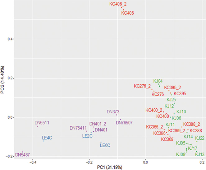
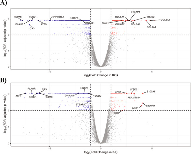
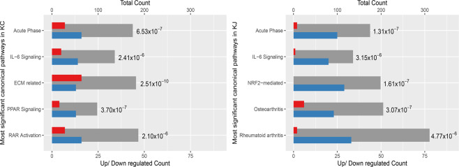
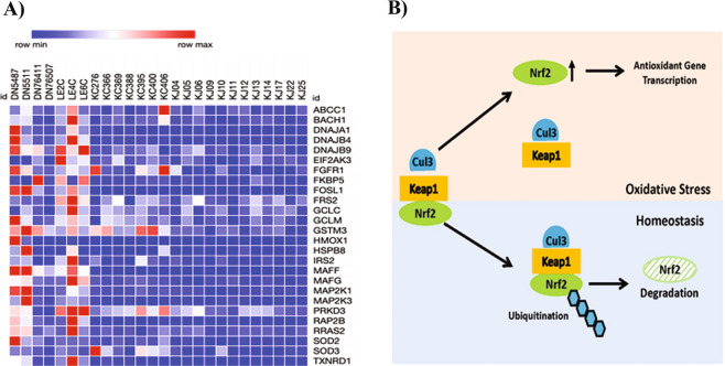
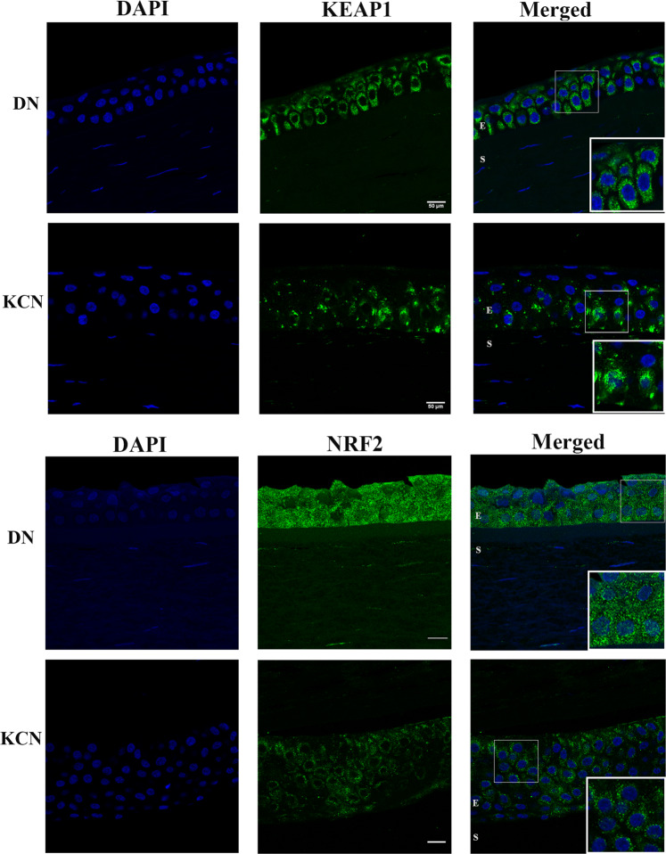
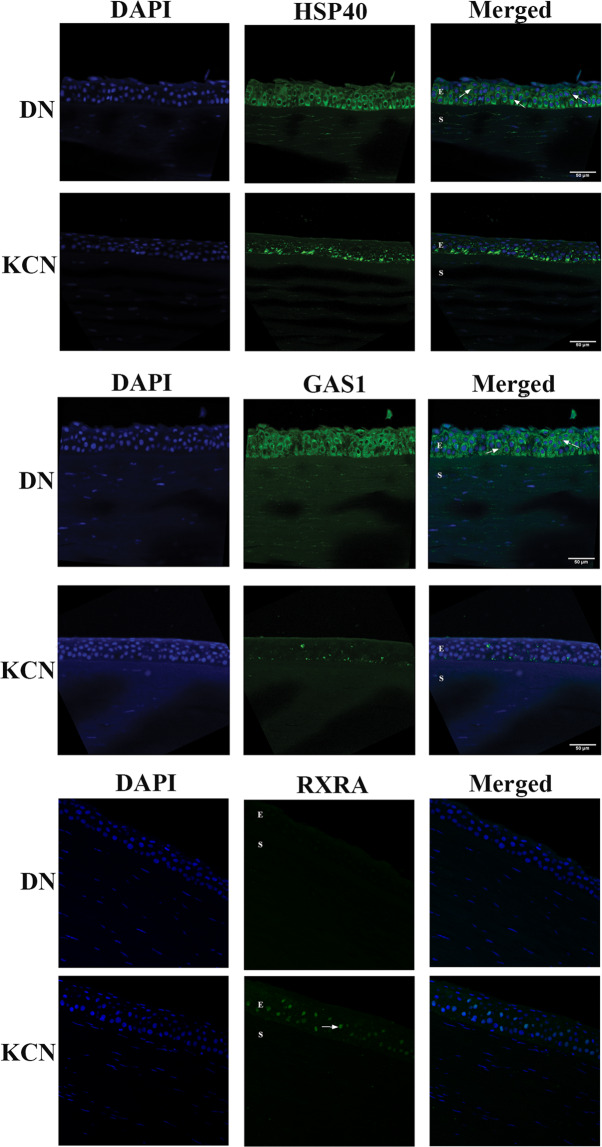
Keratoconus is a highly prevalent (1 in 2000), genetically complex and multifactorial, degenerative disease of the cornea whose pathogenesis and underlying transcriptomic changes are poorly understood. To identify disease-specific changes and gene expression networks, we performed next generation RNA sequencing from individual corneas of two distinct patient populations - one from the Middle East, as keratoconus is particularly severe in this group, and the second from an African American population in the United States. We conducted a case: control RNA sequencing study of 7 African American, 12 Middle Eastern subjects, and 7 controls. A Principal Component Analysis of all expressed genes was used to ascertain differences between samples. Differentially expressed genes were identified using Cuffdiff and DESeq2 analyses, and identification of over-represented signaling pathways by Ingenuity Pathway Analysis. Although separated by geography and ancestry, key commonalities in the two patient transcriptomes speak of disease - intrinsic gene expression networks. We identified an overwhelming decrease in the expression of anti-oxidant genes regulated by NRF2 and those of the acute phase and tissue injury response pathways, in both patient groups. Concordantly, NRF2 immunofluorescence staining was decreased in patient corneas, while KEAP1, which helps to degrade NRF2, was increased. Diminished NRF2 signaling raises the possibility of NRF2 activators as future treatment strategies in keratoconus. The African American patient group showed increases in extracellular matrix transcripts that may be due to underlying profibrogenic changes in this group. Transcripts increased across all patient samples include Thrombospondin 2 (THBS2), encoding a matricellular protein, and cellular proteins, GAS1, CASR and OTOP2, and are promising biomarker candidates. Our approach of analyzing transcriptomic data from different populations and patient groups will help to develop signatures and biomarkers for keratoconus subtypes. Further, RNA sequence data on individual patients obtained from multiple studies may lead to a core keratoconus signature of deregulated genes and a better understanding of its pathogenesis.


Conflict of interest statement

The authors declare no competing interests.

Figures

Figure 1
Figure 1
Histology of patient and donor cornea sections. The figure shows similar retention of epithelial layers in DN and KC corneas, only KJ sample showed irregular and thickened epithelial cell layer in H&E staining of paraffin embedded sections. KC: keratoconus cornea from an African American patient; KJ: keratoconus cornea from a Middle Eastern patient; E: Stratified squamous epithelium; B: Bowman’s Layer; S: stroma; K: keratocytes.
Figure 2
Figure 2
Principal component analysis (PCA) of patient and donor samples. PCA was performed on 19 Keratoconus (7 KC and 12 KJ) and 10 control (7 DN and 3 LE) corneas using 10,652 genes with FPKM value ≥ 5 in at least one sample. Technical duplicates of 7 KC and 1 DN samples (KC*_2 and DN401_2) appear close together. The major separation along PC1 was between KCN and controls. KC and KJ samples cluster together along PC1. African American donor (DN) and KC patient samples are labeled purple and red, respectively; Caucasian donors (LE) are in blue and Middle Eastern patients (KJ) in green. Euclidean distance between samples in a pairwise comparison of samples in PC1 and PC2 is shown in Supplemental Table S3.
Figure 3
Figure 3
Differentially expressed genes (DEGs) in patient groups. (A) DEGs in KC. (B) DEGs in KJ. Among successfully tested genes, 8,762 and 8,990 genes have log2 (fold change) between −8 and 8 in KC and KJ, respectively. Down-regulated genes (q value ≤ 0.01, and fold change ≤−2, and FPKM ≥5 in control or patient groups) are labeled in blue; up-regulated genes (q value ≤ 0.01, and fold change ≥2, and FPKM ≥5 in control or patient groups) are labeled in red; genes not significantly changed are in grey.
Figure 4
Figure 4
Canonical pathways significantly altered in KC and KJ. Grey bars show total number of genes in the pathway (scale on top X axis), with blue and red bars showing the numbers of down-regulated and up-regulated genes (scale on bottom X axis). The p value next to each bar was calculated for that pathway by the IPA Core Analysis. “Acute Phase” is the Acute Phase Response Signaling pathway; “NRF2-mediated” is NRF2-mediated Oxidative Stress Response pathway; “ECM related” is the Hepatic Fibrosis / Hepatic Stellate Cell Activation pathway; “Rheumatoid Arthritis” is the Role of Macrophages, Fibroblasts and Endothelial Cells in Rheumatoid Arthritis pathway; and, “Osteoarthritis” includes genes associated with osteoarthritis.
Figure 5
Figure 5
NRF2 target genes significantly altered in KCN corneas. (A) Decreased NRF2 target gene expression in KC and KJ RNA seq. (B) Mechanism of NRF2 regulation by KEAP1; the latter binds to NRF2 and CUL3 for NRF2-ubiquitination and degradation to maintain low levels of NRF2 under homeostatic conditions. Under oxidative stress, KEAP1 dissociates from NRF2 allowing its increase and upregulation of target genes.
Figure 6
Figure 6
KEAP1 and NRF2 immunostaining in DN and KCN corneas. (A) KEAP1 staining is decreased in KCN corneas, with focally increased staining in some basal epithelial cells (inset), whereas in DN corneas KEAP1 shows staining of all epithelial layers (inset). (B) NRF2 shows very little to no staining of KCN corneas and these were all cytoplasmic (inset), while DN sections show stronger staining of epithelial cells and some nuclear staining (inset) DAPI nuclear staining shown in blue. IF staining of additional KCN and DN cornea sections are shown in Supplemental Fig. S4. Scale bar: 50 µm.
Figure 7
Figure 7
Immunofluorescence staining of selected markers in corneal sections. (A) DNAJA1/Hsp40 is weaker in the KCN, with some staining of basal epithelial cells and more uniform staining of all epithelial layers (E) in DN (arrows). (B) GAS1 staining was almost non-existent in KCN but show uniform staining of all epithelial layers (arrows) and weak stromal (S) staining in DN. These represent one of two KCN and DN cornea samples. (C) RXRA showed a dramatic increase in nuclear staining in epithelial layers of KCN corneas Scale bar: 50 µm.

References

    1. Klintworth GK, Damms T. Corneal dystrophies and keratoconus. Curr. Opin. Ophthalmol. 1995;6:44–56. doi: 10.1097/00055735-199508000-00009. - DOI - PubMed
    1. Krachmer, J. H., Feder, R. S. & Belin, M. W. Keratoconus and related noninflammatory corneal thinning disorders. Surv Ophthalmol28, 293-322, 0039-6257(84)90094-8 [pii] (1984). - PubMed
    1. Bron AJ. Keratoconus. Cornea. 1988;7:163–169. doi: 10.1097/00003226-198803000-00001. - DOI - PubMed
    1. Rabinowitz YS. Keratoconus. Surv. Ophthalmol. 1998;42:297–319. doi: 10.1016/S0039-6257(97)00119-7. - DOI - PubMed
    1. Soiberman U, Foster JW, Jun AS, Chakravarti S. Pathophysiology of Keratoconus: What Do We Know Today. open. Ophthalmol. J. 2017;11:252–261. doi: 10.2174/1874364101711010252. - DOI - PMC - PubMed

Publication types

MeSH terms